Retrobulbar hemorrhage
Retrobulbar hemorrhage - compartment syndrome in the orbit
Postoperative bleeding within the soft tissues of the posterior orbit is termed retrobulbar hemorrhage. This can result in an ischemic and compressive optic neuropathy leading to a loss of vision or damage to other soft-tissue structures.
Detailed postoperative monitoring of vision with documentation is highly recommended. Decompressive procedures for the orbit and optic nerve may be necessary as indicated by declining vision acuity or excessive compartment pressure.
The clinical personnel looking after the patient must be familiar with the typical signs and symptoms of a retrobulbar hemorrhage, optic neuropathy, and orbital compartment syndrome (monitored by tissue pressure).
The patient's head should be elevated postinjury or postoperatively (30°–45°). It is wise to delay fracture treatment until the hemorrhage and compartment syndrome have resolved. Decompressive procedures and the use of sinus or orbital drainage may prevent the occurrence of a retrobulbar hematoma.
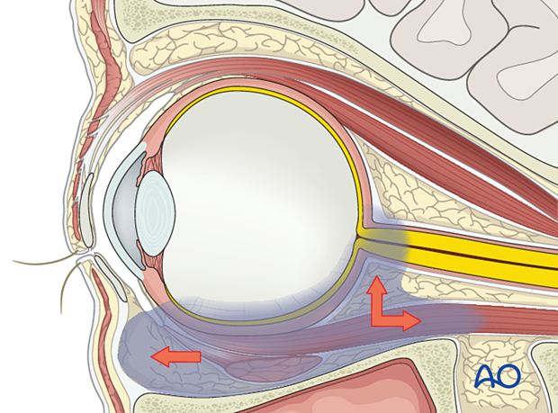
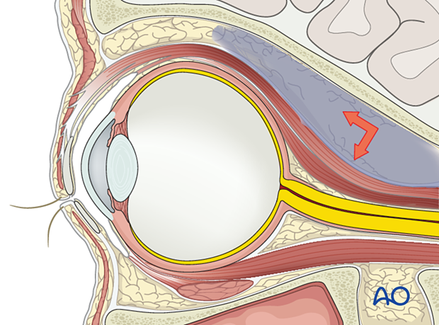
Subperiosteal orbital hemorrhage and compartmentalization of blood within the septal pockets (of the extra or intraconal space) may result in elevation of ocular pressure. This, in turn, may lead to decreased vascular perfusion and ischemia of the optic nerve or the retina, with visual loss as the most ominous complication.
During the intra-orbital dissection, meticulous hemostasis must be achieved to prevent postoperative bleeding. A bloodless field is confirmed prior to insertion of implants or fracture reduction, again after these procedures, and is finally checked again at the end of the intraorbital procedure before closure.
Postoperatively the vision must be monitored regularly.
The signs and symptoms of a retrobulbar hematoma may include:
- Painful proptosis
- Increased orbital tissue tension, increased intraocular pressure
- Ecchymosis of eyelids
- Chemosis
- Decreased visual field
- Decreased visual acuity/loss of vision
- Afferent pupillary defect (APD in swinging flashlight test)

If circumstances do not dictate an emergent intervention, CT or MRI imaging greatly aids in identifying the location and extent of the hemorrhage to plan the decompression and evacuation.
The surgical procedure can be differentiated in an extraconal hematoma of the superior orbit, in contrast to a hemorrhage involving the extra- and intraconal compartments in the lower circumference of the anterior and midorbit regions, as shown in the previous illustration.

Treatment requires prompt intervention with immediate decompression of the expanding hematoma by release of the lateral canthus (lateral canthotomy) and then detachment of the lower limb of the lateral canthus and incision of the orbital septum which provides complete disinsertion of the lower lid from the lateral canthus (inferior cantholysis). This will allow the globe to prolapse freely anteriorly, releasing the compartment pressure within the orbit.
Confined subperiosteal blood collections may necessitate surgical evacuation under direct vision and even decompression of the bony cavity.
Mega dose systemic corticosteroid treatment and osmotic agents are also part of established protocols for treating this condition. The efficacy of corticosteroid treatment and osmotic agents in the treatment of a retrobulbar hematoma or optic nerve injuries has been debated.


