Pre-/Transcaruncular approach
1. Principles
The medial orbital wall can be exposed using a medial conjunctival incision posterior to the lacrimal drainage system along the semilunar fold. The incision is either precaruncular or transcaruncular.
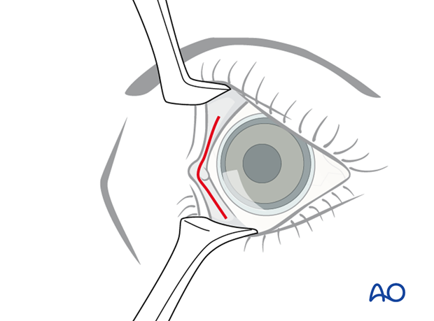
The dissection pathway is continued inside the periorbital soft tissues until the posterior lacrimal crest is reached. The posterior limb of the medial canthus as well as the lacrimal system is left intact. Along the posterior lacrimal crest, the periorbita is incised and the medial orbital wall is dissected directly on the bony surface.
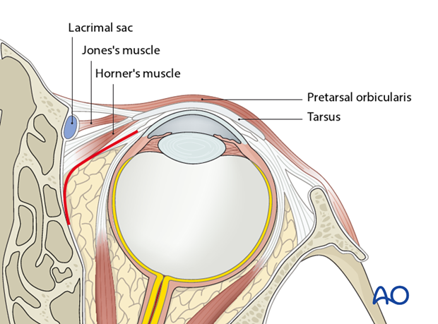
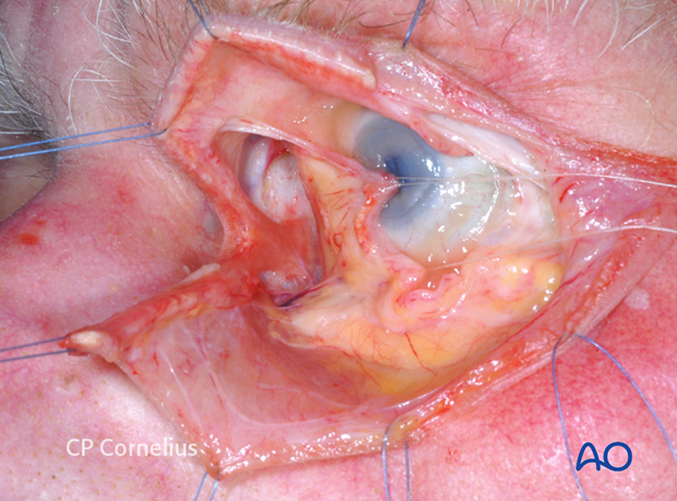
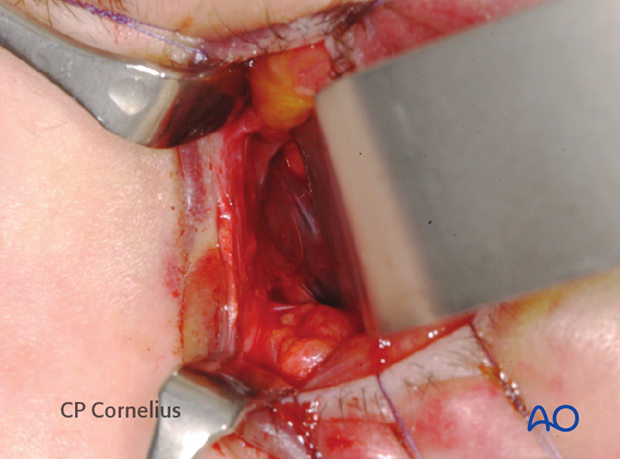
Anatomic specimen showing the precaruncular incision (caruncula grasped with forceps) and the dissection pocket inside the periorbita medially to Horner's muscle. For better visualization the lower lid has been incised and reflected.
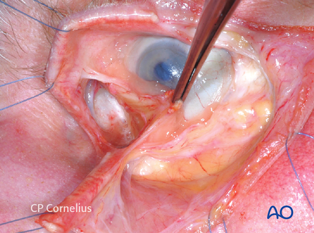
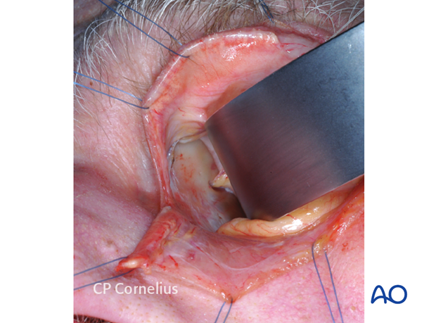
Retraction of the lateral conjunctival edge to fully show Horner's muscle.
After vertical incision of the periorbita posterior of the insertion of Horner's muscle, the medial orbital wall is exposed. The retractor is located just in front of the anterior ethmoidal artery.
2. Vasoconstriction
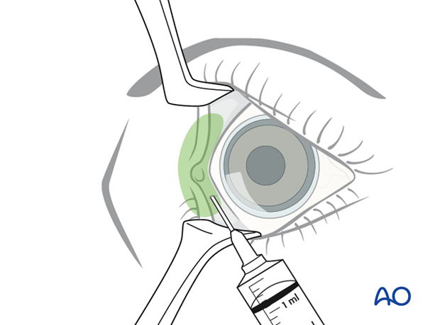
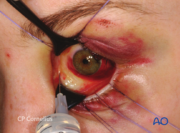
The conjunctiva in the area of the semilunar fold and the caruncle may be infiltrated with a vasoconstrictor. This is done about 7-10 min prior to the incision so that any distortion of the structures can settle by diffusion of the agent.
3. Incision
The upper and lower eyelids next to the medial angle are retracted with traction sutures. Damage to the lacrimal puncta and canaliculi should be avoided. Alternatively, Desmarres or Green retractors can be used.
Additionally, the globe can be moved laterally with malleable Jaeger lid plate retractor or broad spatula.
These maneuvers flatten the caruncle and improve visibility of the incision area.
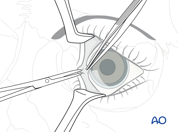
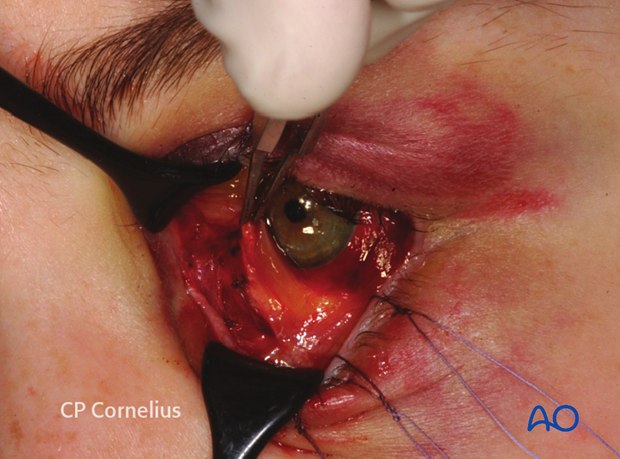
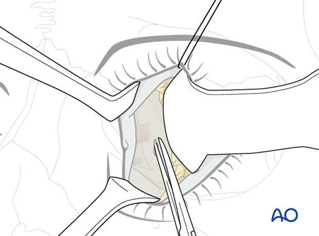
The initial incision with scissors is placed pre- or transcaruncular while the caruncle is grasped with a delicate forceps and pulled laterally.

4. Subconjunctival dissection
Gradually, the incision is extended vertically over a distance of 12-15 mm through the conjunctiva.
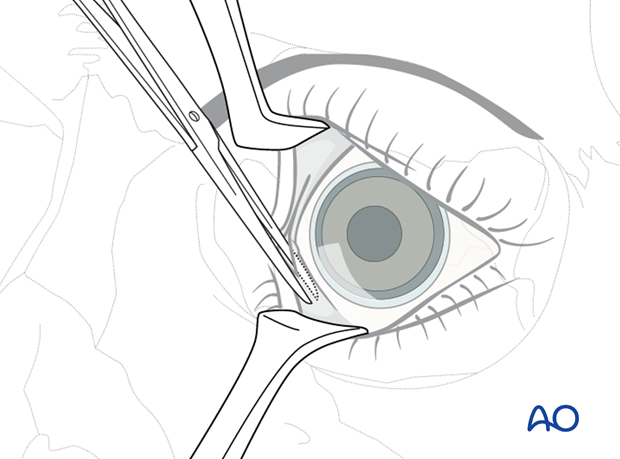
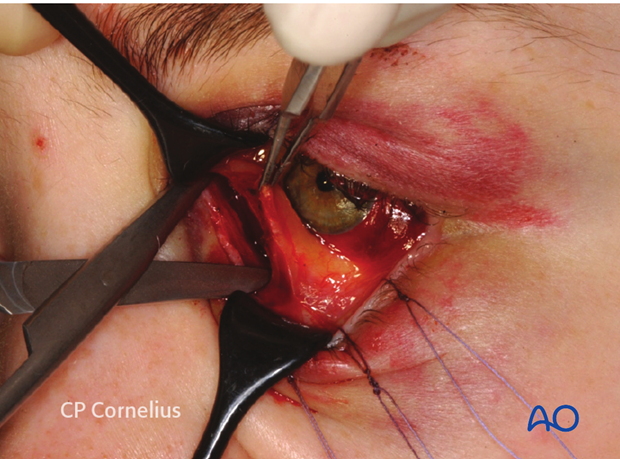
The soft-tissue space deep to the caruncle is spread in a posteromedial direction on top of Horner's muscle. Following the surface of the muscle the dissection will proceed directly to the posterior lacrimal crest where the muscle inserts. The tip of the scissors may be used to palpate the underlying bone.
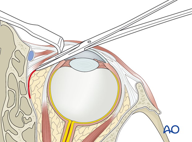
A malleable retractor or periosteal elevator is inserted and directed against the medial orbital wall behind the posterior lacrimal crest.

5. Periosteal incision and exposure
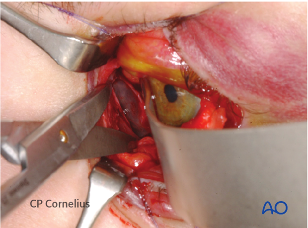
The periorbita along the posterior lacrimal crest is incised in a superoinferior direction with a spreading motion of sharp, pointed scissors.
6. Subperiosteal dissection and exposure of medial orbital wall
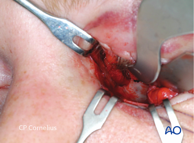
Subperiosteal dissection begins with periosteal elevation. The retractor or periosteal elevator is gradually inserted into the pocket along the bony surface. The medial orbital wall is exposed with sweeping motions from superior to inferior in order to obtain a wide entrance. The ethmoidal arteries indicate the superior extent of the surgical cavity while the inferior extension is limited only by the retractability of the globe.

Clinical example of exposure of the medial orbital wall using a precaruncular approach.

7. Closure
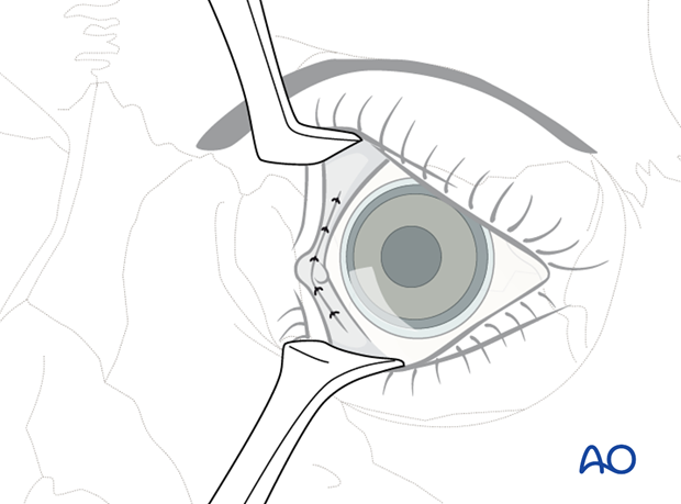
The periorbita is usually not closed. The conjunctiva and the caruncular area are sutured using 6-0 resorbable sutures, either interrupted or partially uninterrupted. The suture ends may be buried.
