Lateral eyebrow approach
1. General considerations
The lateral eyebrow approach provides simple and rapid access to the superolateral orbital rim. No functionally important neurovascular structures are at risk in this approach. As long as the incision is placed within the eyebrow hairs the resulting scar is usually well concealed unless postoperative hair loss is occurring.
A lateral or inferior extension of the incision line outside the hair-bearing portion in order to follow the orbital margin will result in apparent scaring.
Cosmetic eyebrow removal forbids this type of incision for women.
The access through this approach is limited and this previously popular lateral eyebrow incision is rarely indicated except in the East Asian patient. For instance, it may be justified in fractures located high on the lateral orbital rim.
2. Vasoconstriction
Subcutaneous infiltration of a local anesthetic/vasoconstrictor solution of the soft tissues over the superolateral lateral orbital rim is helpful for hemostasis.
3. Protection of the Globe
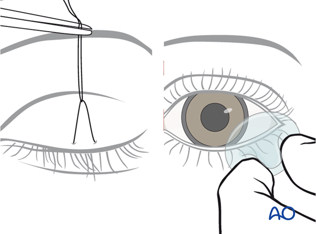
The cornea is protected with a temporary tarsorrhaphy (click here for a detailed description of coronal protection) or with a transparent corneal cover shield allowing undisturbed observation of the eye during surgery.

4. Skin incision
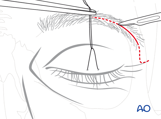
Prior to the incision the hair of the lateral eyebrow is moistened and parted to open a line for the planned incision. An approximately 2 cm long horizontal incision is marked within the bounds of the lateral eyebrow parallel to the hair follicles. The incision goes through the skin first and then through the subcutaneous fat and muscular tissue layers.
Care is taken not to injure the hair follicles during the incision.
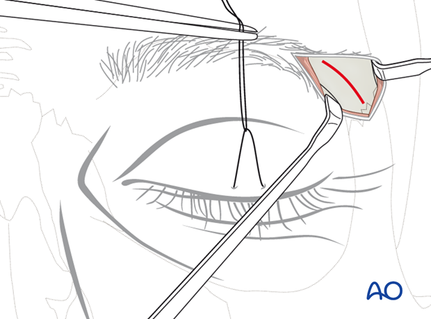
The orbicularis oculi muscle is undermined at a level below the retro-orbicularis oculi fat to expose the supraperiosteal plane.
The wound edges become freely moveable by the supraperiosteal dissection and are retracted over the frontozygomatic suture or the fracture area.

The access area can be enlarged in two fashions:
- Medial extension of the incision towards the supraorbital foramen and nerve staying inside the eyebrow.
- Extending the incision inferiorly along the orbital rim by way of a small angled skin-only turn into a crow's foot wrinkle laterally. Such extensions are positioned at least 6-7 mm above the level of the lateral canthus.
5. Periosteal incision
After exposure of its surface, the periosteum is now split sharply along the middle of the superolateral orbital rim with a scalpel.

6. Subperiosteal dissection of superolateral orbital rim and internal orbit quadrant
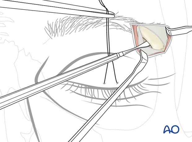
The underlying bony structures are freed using sharp periosteal elevators.
The medial surface of the superolateral orbital rim is exposed first to gain entrance to the fossa of the lacrimal gland. Starting the subperiosteal dissection from within this bony concavity inside the anterolateral portion of the orbital roof, the periosteal envelope can be stripped off with ease from the superolateral orbital rim and the directly adjacent intraorbital quadrant.
Exposure to the lower portion of the lateral orbital rim is facilitated by wide undermining of the skin and periosteum, since this improves the tissue retraction.

7. Closure
Wound closure is in three layers (periosteum, subcutaneous connected tissue, skin).


