Indirect approaches to the zygomatic arch (temporal and transoral approaches)
1. General considerations
The only direct exposure to the zygomatic arch is through a coronal incision. (Click here for a detailed description of the coronal approach).
The coronal incision allows for excellent exposure of the zygomatic arch, as well as reduction and fixation of comminuted fractures. This allows reconstruction of the width and AP projection of the midface. The width at the zygomatic arch defines the maximal width of the face. Proper alignment of the zygomatic arch facilitates definition of the AP projection of the zygoma.
However, the coronal incision results in significant scar alopecia and can also be associated with complications of temporal hollowing and possible risk of injury to the temporal branch of the facial nerve. In many instances, the negative sequelae of the coronal approach are worse than the benefit achieved from direct exposure of the arch. As a result, many surgeons prefer indirect exposures for reduction of the zygomatic arch.
Indirect approaches
Common indirect approaches for reduction of the zygomatic arch include:
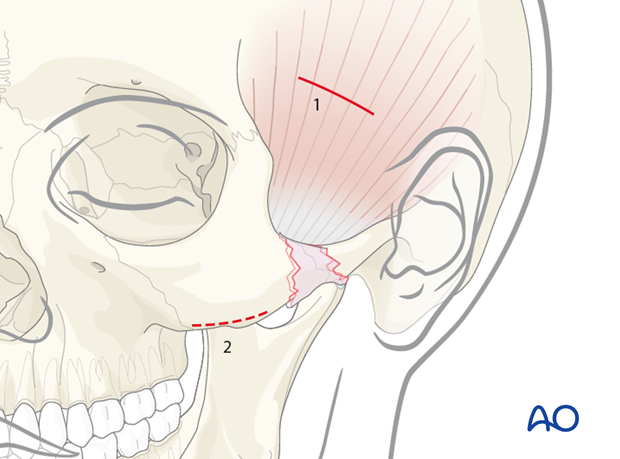
- Temporal (Gillies) approach (1)
- Transoral (Keen) approach (a lateral maxillary vestibular incision), (2)
An impact to the lateral side of the face can sometimes result in a pure zygomatic arch fracture, where the zygomatic complex itself remains nondisplaced. In this case, two open approaches can be considered for a reduction of the zygomatic arch: the temporal (Gillies) and transoral (Keen) approach.
2. Temporal (Gillies) approach - Skin incision
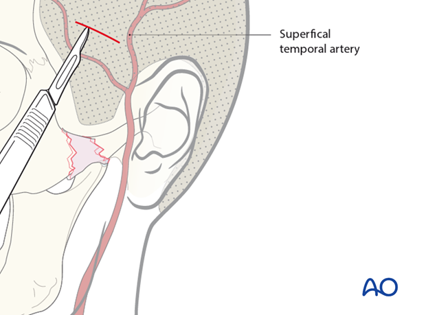
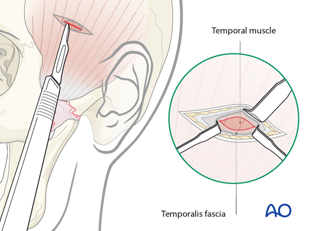
The Gillies technique describes a temporal incision (2 cm in length), made 2.5 cm superior and anterior to the helix, within the hairline.
A temporal incision is made. Care is taken to avoid the superficial temporal artery.
Temporal (Gillies) approach - Deep dissection
The dissection continues through the subcutaneous tissue and superficial temporal fascia down to the deep portion of the deep temporal fascia.
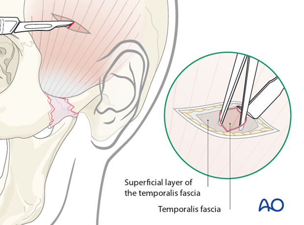
This fascia is then incised to expose the temporalis muscle.
Temporal (Gillies) approach - Exposure
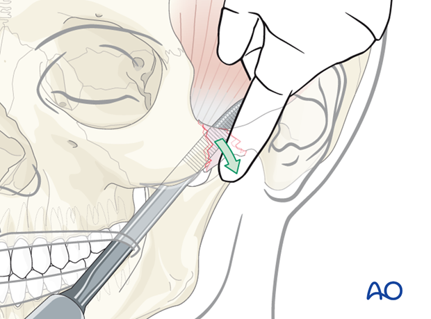
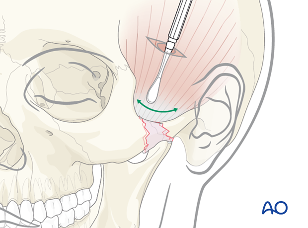
An instrument is inserted deep to the temporalis fascia and superficial to the temporalis muscle. Using a back-and-forth motion the instrument is advanced until it is medial to the depressed zygomatic arch.
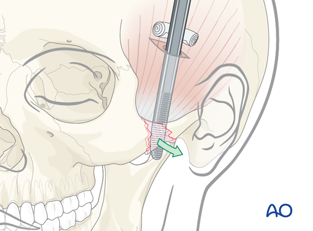
A Rowe zygomatic elevator is inserted just deep to the depressed zygomatic arch and an outward force is applied.
Great care should be taken not to fulcrum off the squamous portion of the temporal bone.
Temporal (Gillies) approach - Wound closure
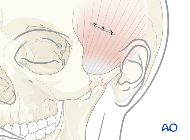
Closure is performed according to the preference of the surgeon.
3. Transoral (Keen) approach – lateral maxillary vestibular incision
The transoral (Keen) approach provides the most direct access to the zygomatic arch.
It allows for an intraoral incision, and therefore does not have the risk of scar alopecia that will result from a temporal (Gillies) approach.
A 2 cm lateral maxillary vestibular incision (upper gingival buccal incision) is made with a scalpel or a cautery device just at the base of the zygomaticomaxillary buttress. The incision is made through mucosa only.
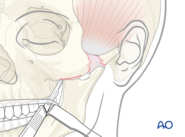
Transoral (Keen) approach - Exposure
Because of the direct proximity of the incision to the arch, an instrument can easily be placed deep to the fractures to allow elevation of a depressed zygomatic arch. Because of the direct proximity of the incision to the arch, the depressed arch can often be palpated and elevated with a digital exam.
