Approaches to the maxilla
1. Overview
Relevant to the required exposure in trauma of the midface two different approaches are possible:
2. Maxillary vestibular approach
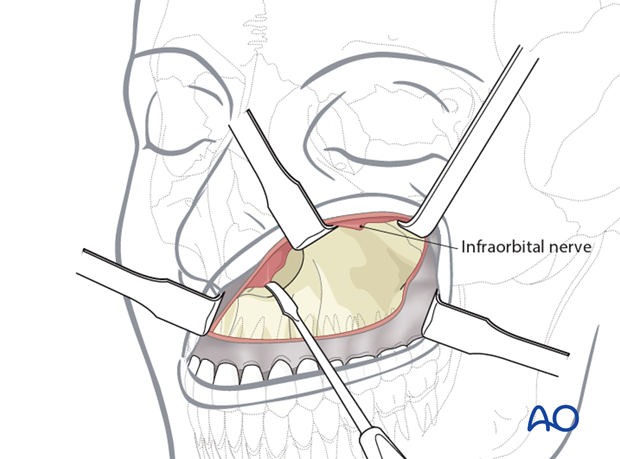
In the surgical treatment of facial trauma, the skeleton of the lower midface is commonly exposed using a transoral approach in the maxillary vestibule.
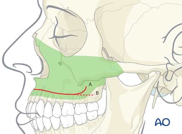
A horizontal incision (possibly with hockey stick endings) through the maxillary vestibular mucoperiosteum is made slightly above the mucogingival junction. The subsequent subperiosteal dissection can be advanced to access the entire anterolateral bony surfaces from the zygomaticotemporal suture over the facial antral wall to the borders of the piriform aperture and up to the infraorbital rim including the infraorbital nerve exit.
Click here for a detailed description of the maxillay vestibular approach.

Access area
The anterolateral surfaces of the lower midface skeleton can be exposed via a unilateral or bilateral maxillary vestibular approach:
- Entire anterior face of the maxilla
- Zygomaticomaxillary buttress
- Infraorbital rim
- Zygomatic body, anterocaudal part of zygomatic arch
- Piriform aperture
- Anterior nasal spine and caudal nasal septum
Neither the posterior wall of the maxilla or the pterygomaxillary fissure and fossa can be directly visualized.
The length of the vestibular incision and extent of subperiosteal dissection are varied according to the area of surgical interest and the extent of the intervention. In case of a zygomaticomaxillary fracture, the incision is limited to one side.

3. Midfacial degloving approach
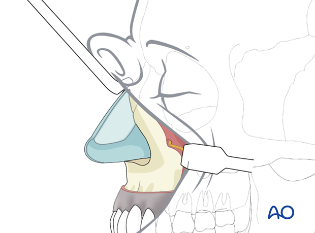
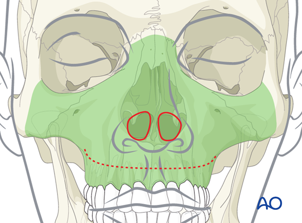
Midfacial degloving is an option rarely used in facial trauma. The horseshoe type bilateral maxillary vestibular incision is combined with a circumferential incision (intercartilaginous, transfixion, and nasal floor) inside both nostrils. This enables lifting the soft-tissue envelope all the way up to the nasal dorsum, radix and ethmoid region.
Click here for a detailed description of the midfacial degloving approach.

Access area
With the circular intranasal incisions and lifting of the midfacial envelope, the access area is extended cranially into the nasal dorsum and ethmoid area as well as the entire zygomatic body and the lower portion of lateral orbital rim.

4. Links to detailed descriptions
Click the following links to read a detailed description of the approaches to the maxilla:


