Dentoalveolar fractures
1. Diagnosis
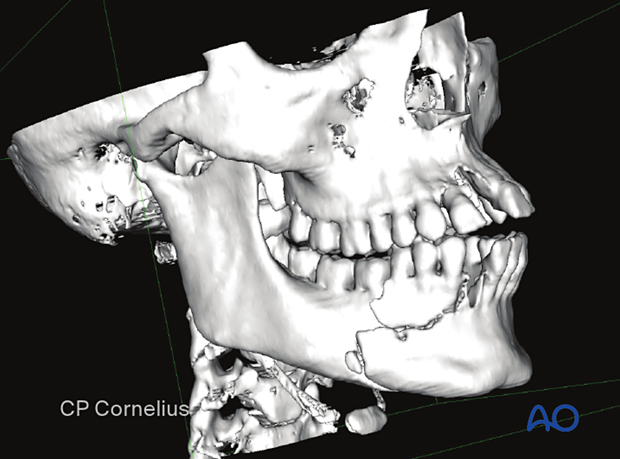
Alveolar fractures can occur both with and without the involvement of the basal bone. In this case, both are present.
Routine diagnosis of alveolar fractures should include a CT scan and/or a panoramic radiograph. Periapical and occlusal dental x-rays can be beneficial.
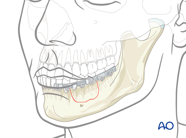
The fracture of the alveolar process reaching from the canine to the first molar is visible on this 3D CT.

2. Treatment options
Alveolar process fractures can usually be treated by reduction and fixation with an arch bar that must be maintained for approximately six weeks to provide time for the fracture to heal.
As an alternative, open reduction and internal fixation may be used in selected isolated alveolar fractures and mostly in those associated with more severe mandibular fractures. Sufficient size of the teeth-bearing bone fragments is required to position the miniplates and screws without damaging the dental roots.
Tooth luxation and fractures are commonly associated. Teeth in a fracture line should be carefully assessed, clinically and radiographically, to determine the need for extraction.

3. Case example
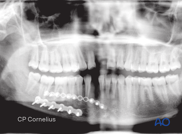
In this case, it was possible to include the alveolar fragment using a miniplate plate fixed with monocortically inserted screws located adjacent to the tooth apices. The mandibular fracture was treated with a large profile locking plate 2.0 to give enough stability along the inferior mandibular border.


