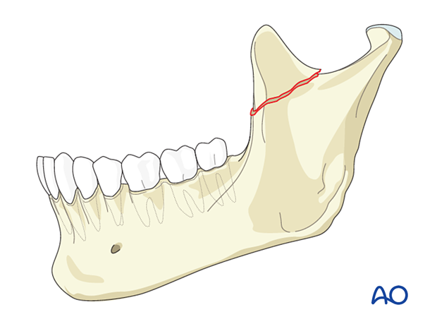
Coronoid process fractures
1. Diagnosis
Fractures of the coronoid process are relatively uncommon. It is generally accepted that they account for approximately 1% of all mandible fractures. The coronoid process is infrequently fractured because the zygomatic arch protects it.
A diagnosed coronoid fracture is an indication for the surgeon to look for an associated zygomatic arch fracture.

Coronoid fractures are often incidental findings on x-rays when looking for associated maxillofacial injuries.
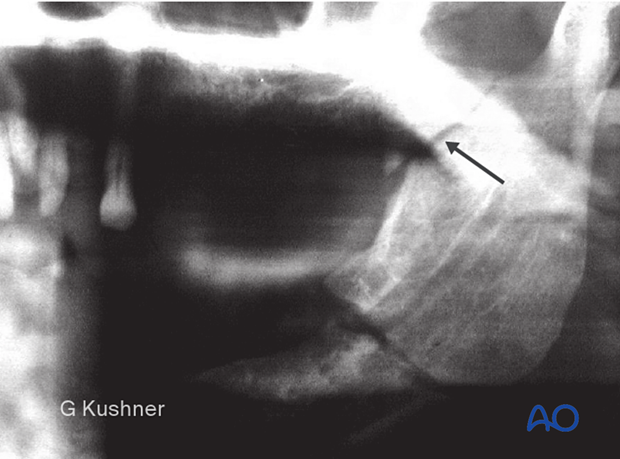
Diagnosis of a coronoid process fracture would most likely be made on a panoramic x-ray or CT scan. Most commonly, there are other associated fractures. Isolated fractures of the coronoid process are extremely rare.

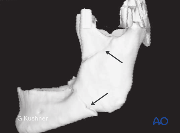
CT scans provide the best delineation of coronoid fracture morphology. This CT scan demonstrates a fracture of the left coronoid process.
Note the associated fracture of the mandibular angle region.

2. Indications for treatment
Very rarely does a fracture of the coronoid process require treatment. Most fractures are observed. Some surgeons would advocate the removal of the coronoid process in case of persistent mouth-opening restrictions.
In a surgically treated comminuted ramus fracture, an associated coronoid fracture may be reduced and fixed.


