Body, simple
Definition
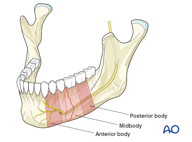
These are single fractures between the mandible canine teeth and the line between the second and third molar.
Images
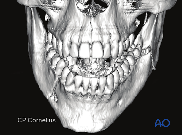
This 3-D reconstruction shows a simple right anterior body fracture with an associated left mandibular angle fracture.
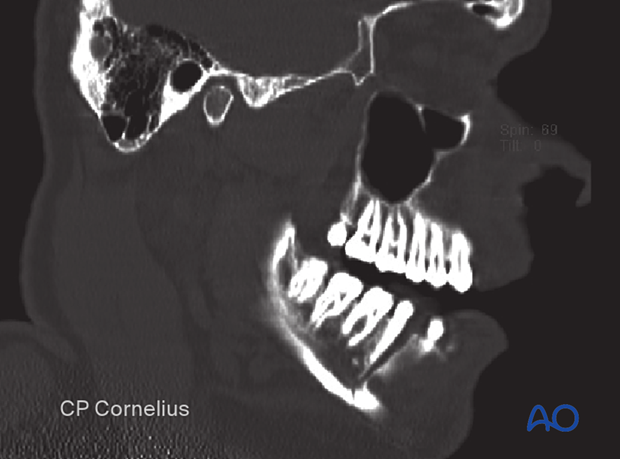
This Sagittal CT scan is of the same patient.
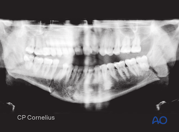
Orthopantomogram shows the same patient with bilateral fractures.
CBCT (cone beam computed tomography) allows for 3-D analysis and reconstruction based on a volumetric data set. In some cases, CBCT allows for a lower radiation dose compared to a CT scan.
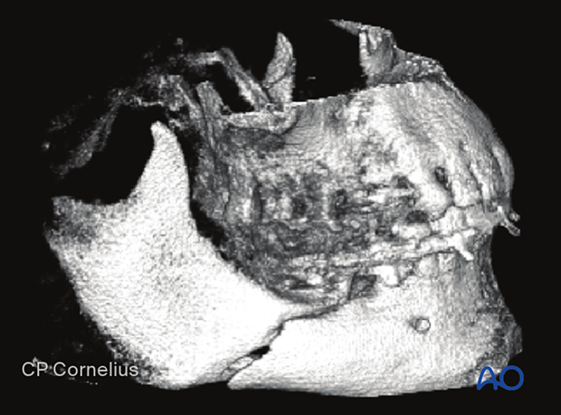
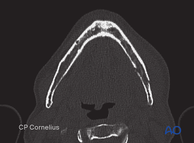
Sagittal split fractures can exist in any part of the mandible. They can best be visualized on the axial CT as an oblique fracture through the axial plane.
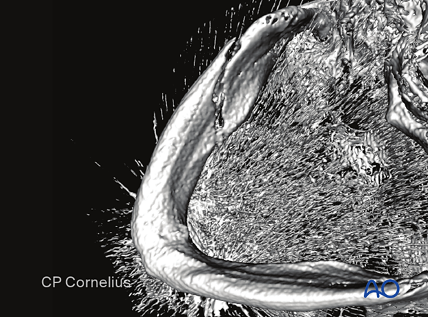
This 3D reconstruction shows a different view of the same sagittal split fracture. The obliquity of the fracture is not evident in this view.
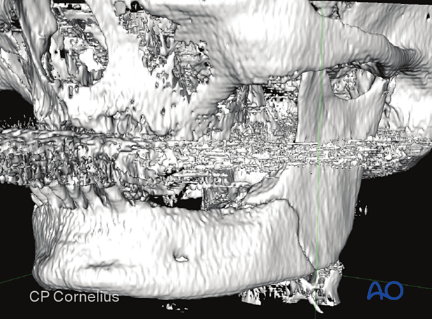
The obliquity of this fracture is again noted in this axial CT.
Mechanism of the injury
Simple body fractures most often result from direct impact during a physical altercation. Fractures of the mandibular body rarely occur in isolation in the dentate mandible. As a general rule, if you suspect a body fracture, also look for a a fracture on the contralateral side.
The frequency of midbody fractures is rare in comparison to the anterior body and posterior body fractures. The latter two locations represent points of weakness due to the biomechanics of the mandible. In the anterior body, the canines' tooth roots and the premolars are responsible for decreased mandibular stability. In the posterior body, the lever relations close to the angle make up for the weakness.
They are usually combined with fractures in the contralateral hemimandible or with ipsilateral fractures in the ramus or subcondylar region.


