Submental approach
1. Principles
The submental approach is used to treat fractures of the anterior mandibular body and symphysis.
Although these fractures are usually approached and treated intraorally, a submental approach may be indicated. Its indication will depend on the fracture severity, and/or the presence of a laceration.
An advantage of this approach is that the surgeon can easily inspect the mandible's lingual surface to assure optimal reduction.
There are no major neurovascular structures in the submental area.
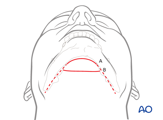
The following variations can be used
A) Following the curvature of the anterior mandible
B) Hidden in the submental skin crease
According to the anatomy and surgical preference, both techniques offer adequate access to the submental region.
2. Skin incision
Use of vasoconstrictors
The use of a solution containing vasoconstrictors ensures hemostasis at the surgical site. The two options currently available are the use of local anesthetic or a physiologic solution with vasoconstrictor alone.
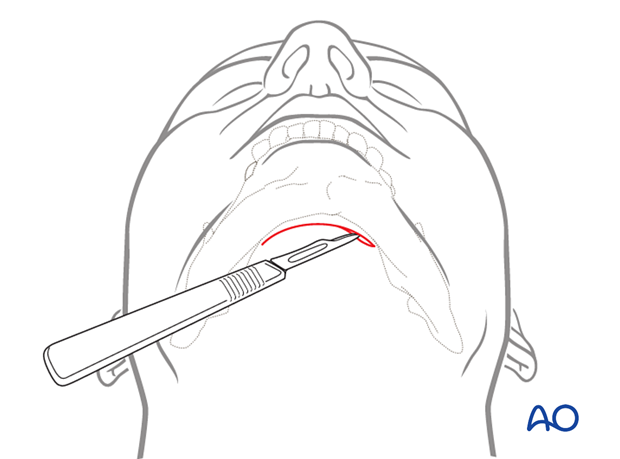
Skin incision
Incise the skin along the selected path.
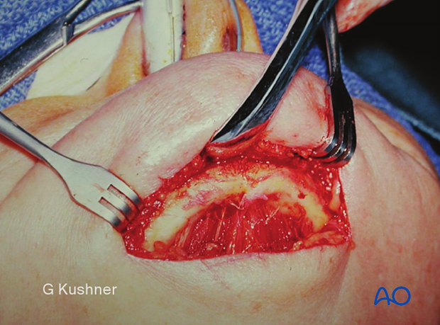
3. Dissection
Carry the incision through the skin and subcutaneous tissues to the platysma muscle.
The platysma muscle must be divided in the midline.
There may be a natural separation of the muscle in the midline region. Additionally, the platysma muscle can become very thin in this region.
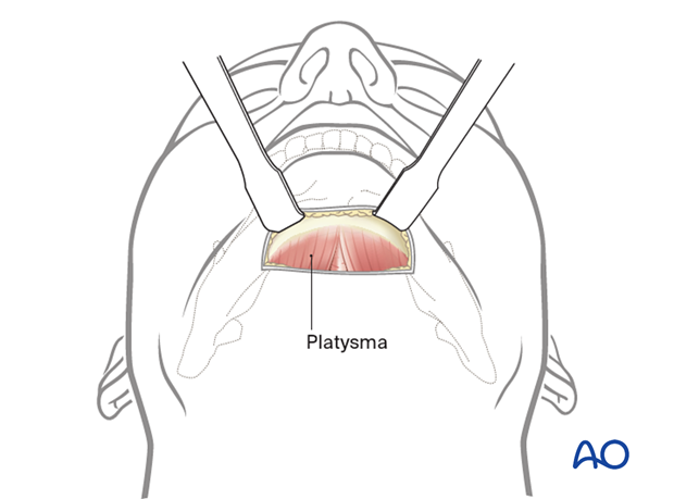
Dissection is carried out to the inferior border of the mandible. The periosteum is incised sharply, and the flap is elevated to expose the anterior surface of the symphysis.

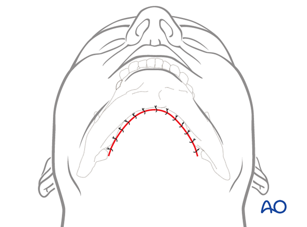
4. Wound closure
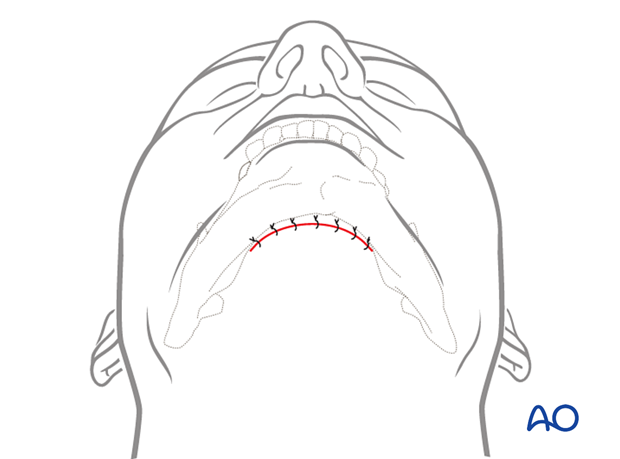
The wound is closed in layers to realign the anatomic structures and to eliminate dead space.
The periosteum and platysma muscle should be closed in different layers.
A variety of skin closure techniques are available according to surgical preference.
5. Option: bilateral extension
Submental extension
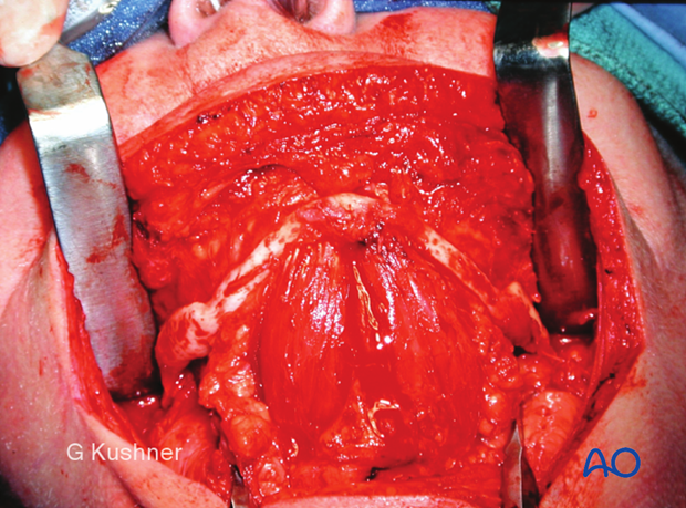
The submental incision can be extended laterally to encompass both the right and left mandibular body. The entire lateral surface of the mandible is degloved in the same way as in the submandibular approach.
This may be necessary in complex fractures such as comminuted, atrophic, and severe bilateral fractures.
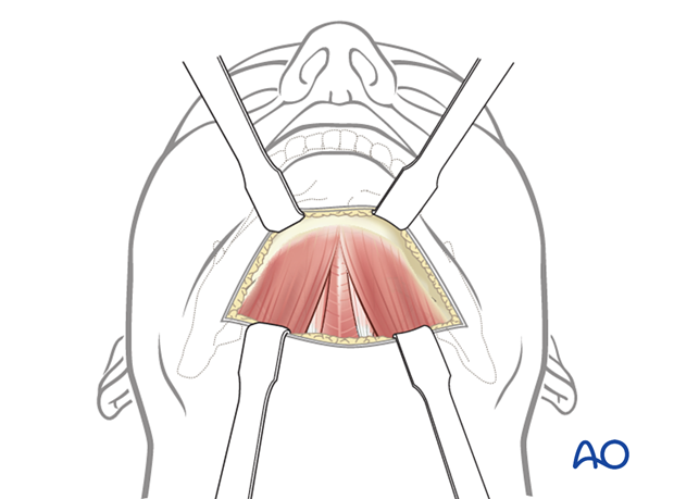
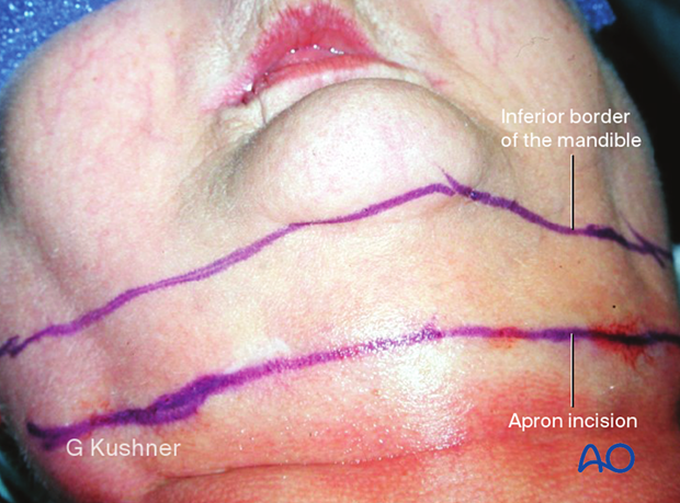
An extension of the incision parallel to the inferior border is shown in the following illustrations as the approach is explained.
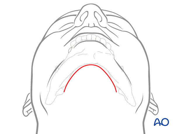
To approach complex mandibular fractures the surgeon essentially combines a right and left submandibular incision with a submental one.
The inferior border of the mandible is marked along with the planned skin incision. These larger incisions are sometimes referred to as an apron incision.
Clinical image of an exposed edentulous atrophic fractured mandible.
Closure
The wound is closed in layers to realign the anatomic structures and eliminate dead space. A variety of skin closure techniques are available based on surgical preference. A drain may be used if necessary.
