Observation
1. Treatment
The patient is assessed clinically at regular intervals by monitoring the occlusion radiographically to assure that union occurs and the fracture does not displace. The patients are maintained on a no-chew diet for several weeks.
2. Case example
Pretreatment imaging
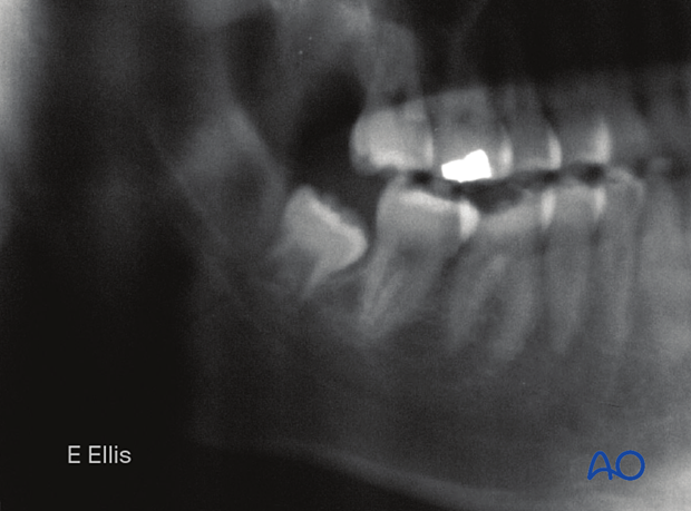
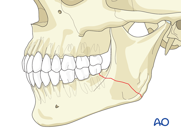
The x-rays show nondisplaced, incomplete, closed fracture through the mandibular angle.
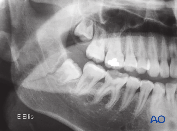
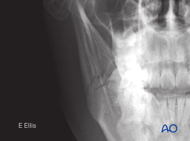
This image shows the PA view of the same patient.
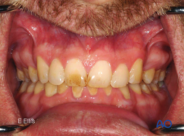
Pretreatment occlusal relationship
The occlusion was normal, and there was no perceptible mobility of the fracture
Outcome
Six weeks later, the x-ray shows that the occlusion is still unchanged (normal), and the fracture has healed.
