Tooth luxation, with displacement - Lateral luxation
Definition and clinical appearance
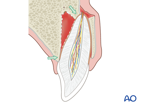
Lateral luxation implies lateral eccentric displacement of the tooth in its socket. It is often accompanied by fracture of the alveolar bone plate.
Clinically, the tooth appears laterally displaced and is generally stable in its new position.
Response to pulp test is generally absent. Exceptions include cases with minimal displacement.

Clinical findings
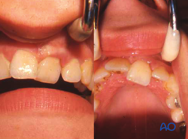
Frontal and palatal view of left central maxillary incisor with lateral luxation. The tooth is only attached to the palatal mucosa, and it is displaced in palatal direction.

Radiographic findings
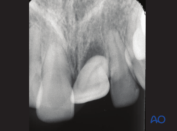
Intraoral film demonstrates that the tooth is axially dislocated out of its socket with partial or total loss of bony attachment.


