Tooth fracture, root fracture
Definition and clinical appearance
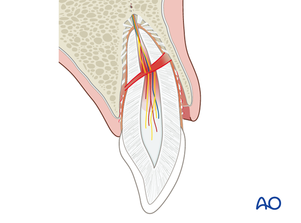
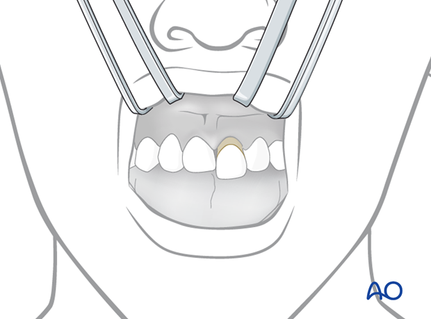
Root fractures are intraalveolar fractures involving dentin, cementum, and the pulp. They are usually combined with extrusion injury of the coronal fragment. The tooth appears elongated. Maxillary teeth are usually displaced palatally.
The tooth is very loose with bleeding from the gingival sulcus.
Response to pulp testing is often absent. Exceptions include cases with minimal displacement.

Clinical findings
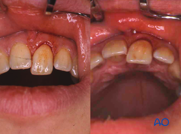
The tooth appears coronally displaced and loose. Occlusal interference is frequent. The condition is clinically indistinguishable from an extrusive luxation.

Clinical photographs showing extrusion of the coronal fragment.

Radiographic examination
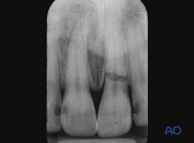
Intraoral films will show the root fracture usually diagonal and located in the apical, middle, or cervical third of the root, or combinations of these (middle cervical). There is normally a fracture gap between the two fragments. The magnitude of the fracture gap is decisive for subsequent wound healing. In optimal situations hard-tissue bridging occurs. In less optimal situations where a hard-tissue bridge is not formed, there will be interpositions of periodontal ligaments between the fragments, resulting in hypermobility of the coronal fragment. Nonhealing may occur due to formation of pulp necrosis located in the coronal fragment which can be managed successfully with endodontic treatment.
The apical fragment is generally nondislocated.


