Pulp capping / partial amputation / extirpation
1. Decision/Indication
Depending on the extent of the exposure, the circulatory status of the pulp, and the stage of root development, three treatment options may be considered:
- Pulp capping
- Partial pulp amputation (pulpotomy, ie, partial removal of pulp)
- Pulp extirpation (removal of entire pulp)
All three methods are highly technique sensitive and should be performed under uncompromised treatment situations where adequate moisture control and sterility can be maintained. Experience has shown that delayed treatment does not compromise the prognosis. Exposed pulp does not normally result in spontaneous pain but pain reaction can be elicited by mechanical stress (chewing), thermal stimulation (exposure to cold and heat), and chemical irritants (acid beverages). It may be tempting to cover the exposed pulp with cement, but experience has shown that this can lead to anaerobic bacteria accumulation under the filling, which is very harmful to the pulp. Therefore coverage with cement should not be performed.
There is a significant window of opportunity for the treatment of crown fractures with exposed pulp. Definitive treatment may be deferred within 1 week with negligible influence on the prognosis.
Pulp capping is a method of compromise which can be performed without cavity preparation in order to preserve the pulp. Capping is especially valuable in teeth with incomplete root formation. Successful pulp capping will preserve pulp vitality and thus allow further root formation in teeth with incomplete root formation.
Prognosis depends on the maintenance of sterility and a tight adhesion of the covering material applied immediately.
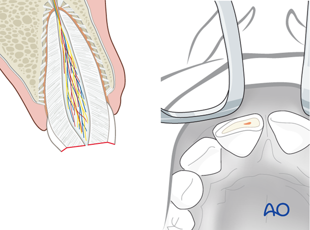
Partial pulp amputation (pulpotomy) is the treatment of choice to preserve the pulp, and especially valuable in teeth with incomplete root formation. Successful pulpotomy will preserve pulp vitality and thus allow further root formation in teeth with incomplete root formation.
Prognosis depends on maintenance of sterility and on tight adhesion of the immediately applied covering material.
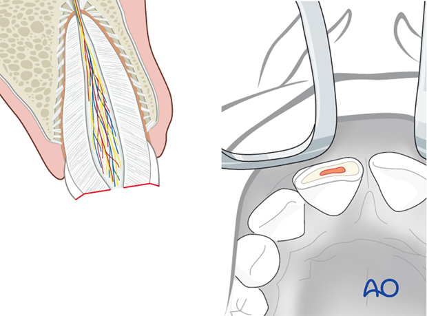
Pulp extirpation is indicated in teeth with fully developed roots and no obvious signs of pulp circulation. A deposit of antibacterial root canal disinfectant may be useful as a preventive measure before final root canal filling and restoration of the tooth, preferably after 1 week.
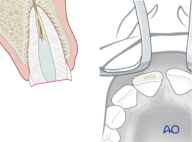
2. Pulp capping
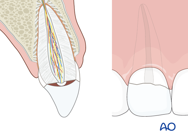
A calcium hydroxide seal or mineral trioxide aggregate (MTA) is first placed to cover the exposed pulp tissue and a suitable provisional filling is added.
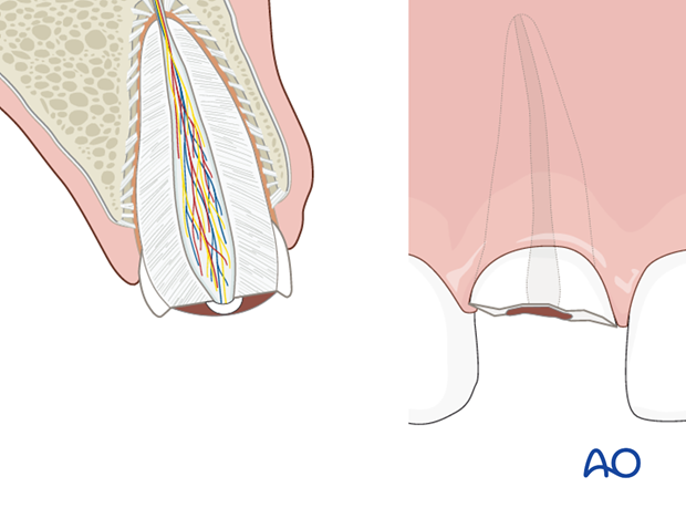
If feasible, the fractured crown segment may be reattached immediately with etch/composite bonding technique.
If reattachment is delayed, the crown fragment should be stored in saline.
3. Pulpotomy
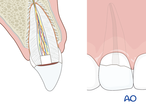
The exposure of pulp may be widened, and the superficial part of the pulp removed using a sharp round burr to a depth of 2–3 mm. A calcium hydroxide seal is placed in the root canal and a suitable provisional filling added.
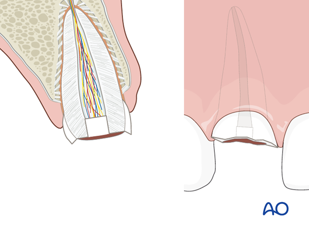
If feasible, the fractured crown segment may be reattached immediately with etch/composite bonding technique.
If reattachment is delayed, the crown fragment should be stored in saline.
4. Pulp extirpation
Principle
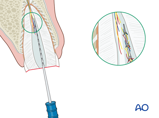
Pulp extirpation aims at the removal of all necrotic pulp tissue. Since the assessment of vitality of pulpal tissue apical to the exposed pulp lesion in a crown fracture is not feasible, an extirpation of all pulpal tissue is advisable leaving 2 mm of apical pulp.
A deposit of antibacterial root canal disinfectant may be useful as a preventive measure before final root canal filling and restoration of the tooth.
To learn more about pulp extirpation go to the Dental Trauma Guide (external link, click here)

Removal of necrotic pulp
Remove the necrotic pulp using an extirpation needle. All pulpal tissue should be removed, leaving 2 mm of apical pulp
Placement of disinfectant and sealing of root canal
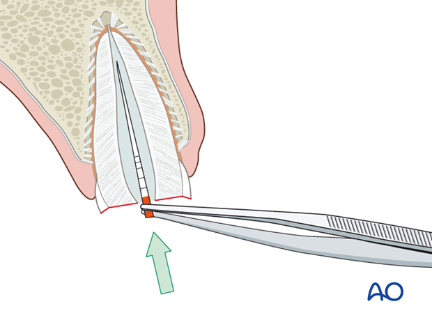
A paper point with a suitable disinfectant is deposited in the root canal until definitive endodontic treatment and root filling can be performed. Alternatively, calcium hydroxide paste may be placed in the root canal. The root canal should then be sealed with a temporary filling.
This completes the acute management of complicated crown fractures with pulp necrosis. Definitive endodontic treatment and restoration of the tooth are left to the general practitioner of dentistry.
5. Aftercare following treatment of enamel-dentin-pulp fractures
Aftercare includes follow-up of pulpal vitality and restoration of the crown. If pulp vitality is retained at 6 months posttrauma no further follow-up is needed. In case of pulp necrosis, appropriate endodontic therapy has to be implemented.


