Tooth fracture, enamel only/dentin exposure
Definition and clinical appearance
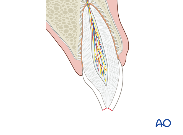
An enamel fracture is confined to the enamel and frequently shows a loss of substance (enamel).
Pulp testing is advisable to ensure pulpal health and for later documentation.
Radiographic findings
In some cases the loss in enamel will be apparent on thex-ray. The main purpose of the x-ray is to diagnose subgingival/submucosal hard-tissue injuries and to exclude preexisting pathology.
A soft-tissue weighted x-ray may reveal radiopaque material (tooth segments, bone, foreign bodies, etc.) implanted in the labial tissues in soft-tissue lacerations.
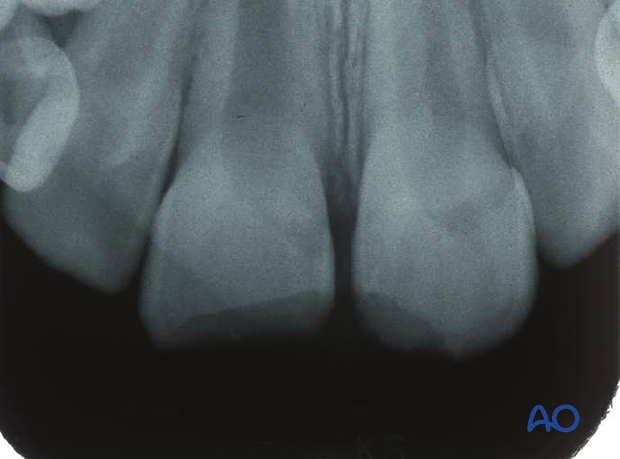
X-ray shows enamel fractures in the two central incisors with limited loss of substance.
X-ray shows tooth fragments in the soft tissues of the lower lip.
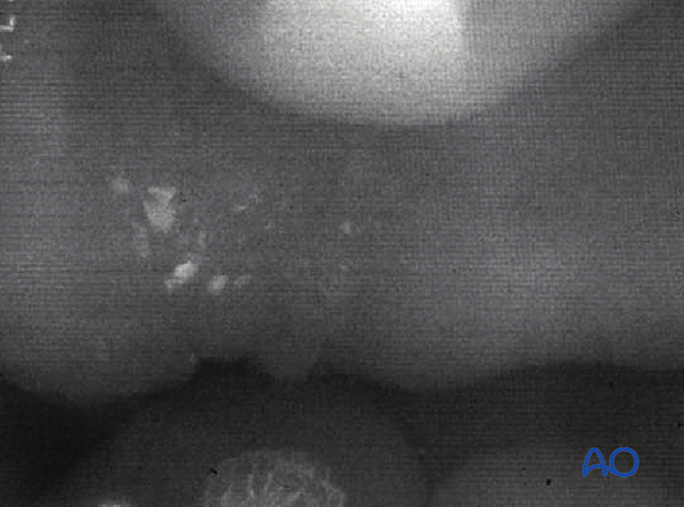
Foreign bodies such as these have to be removed.
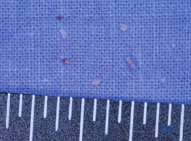
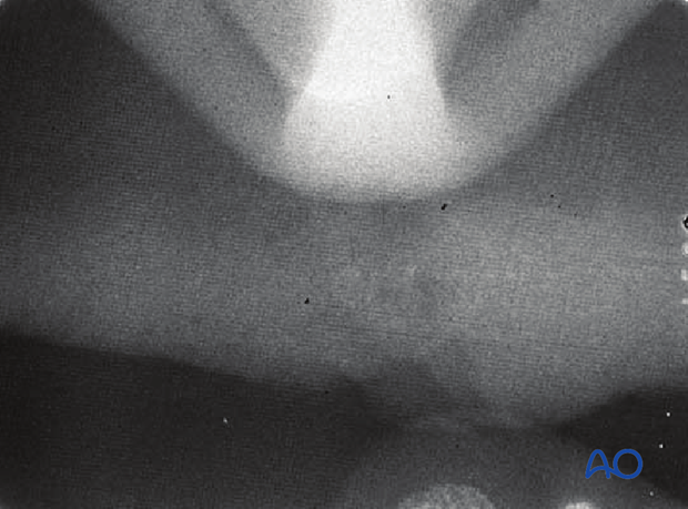
Postoperative x-ray verifying the removal of the radiopaque fragments.
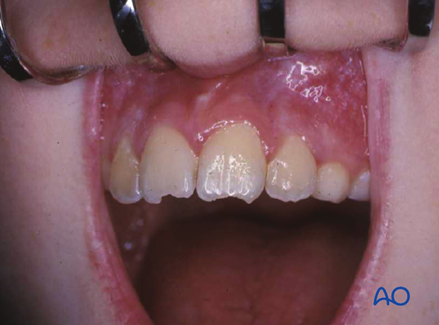
Clinical photographs showing the same case.
Enamel-dentin fracture: Definition and clinical appearance
Enamel-dentin fractures are represented by the loss of tooth substance confined to enamel and dentin, but not involving the pulp.
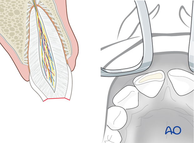
Enamel-dentin fracture: Clinical findings
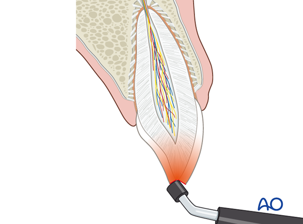
A fragment of a tooth crown is missing. If a major fragment is retained, it should be stored in water, preferably saline, for later reattachment with composite etch technique. The tooth may prove sensitive to temperature change.
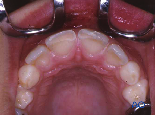
Clinical photographs showing fracture of two incisors with loss of major crown fragments.
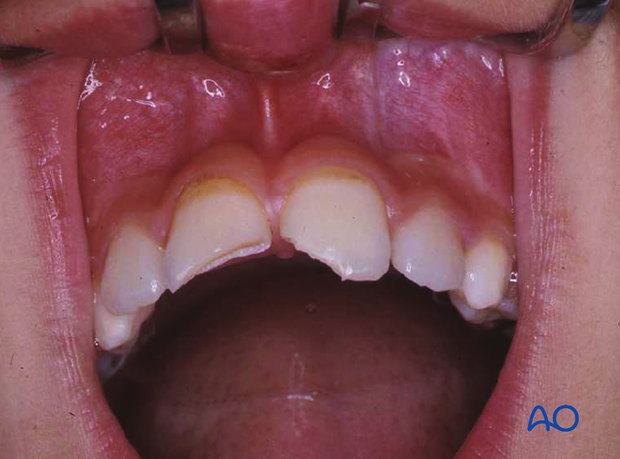
Dentin is exposed but not the pulp.
Enamel-dentin fracture: Radiographic findings
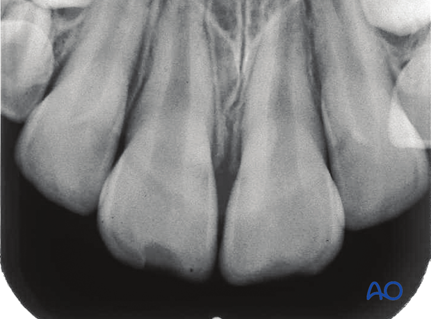
The lost tooth substance is apparent on the x-ray. It is important to ensure that there is no associated root dislocation, root fracture, or other pathology.
