Tooth fracture, crown-root fracture
Definition and clinical appearance
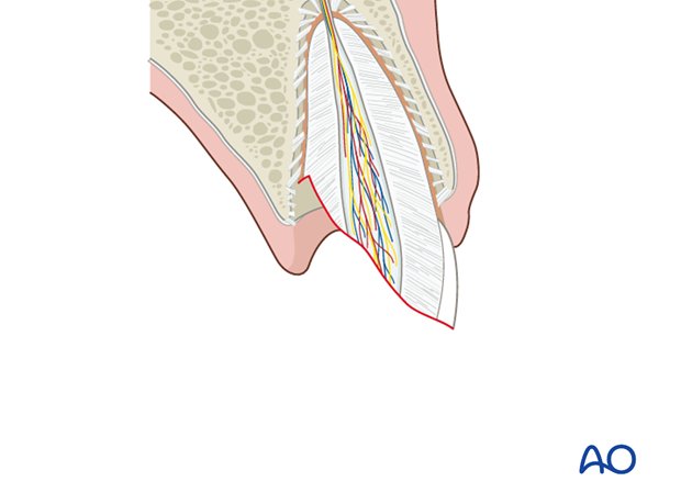
This is a fracture involving enamel, dentin and cementum, without or …
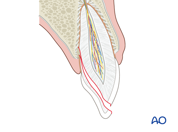
… with pulpal exposure.
In both cases, the fracture by definition crosses the seal of the gingival crevice, thereby involving the coronal part as well as the intraalveolar part of the tooth, thus making this a complex wound.
Clinical findings
The coronal fragment is frequently in-situ and attached to the gingiva.
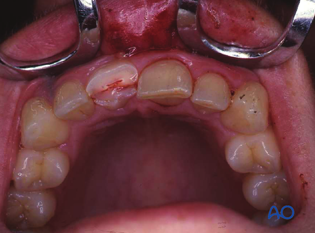
The clinical photograph shows a case where a crown-root fragment has been lost.
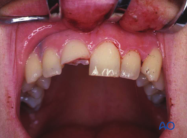
The fracture line may be very obvious but sometimes only presents as a crack. The axial view shows the orientation of the fracture taking a mesiodistal course, and thus can not be seen on the following x-ray.
Radiographic findings
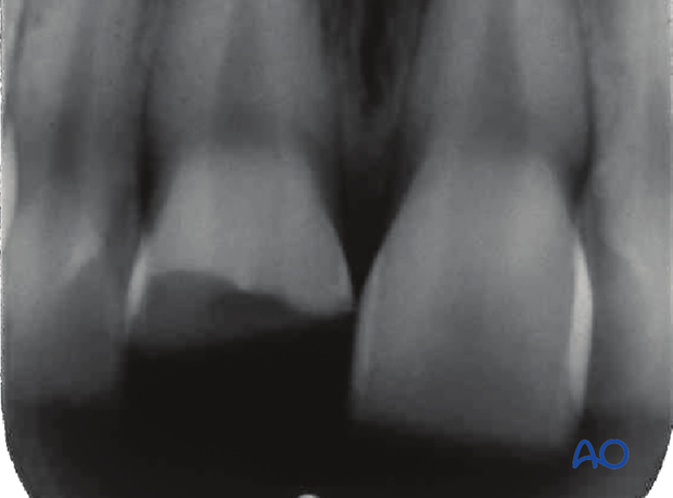
Depending on the orientation of the fracture plane, it may or may not be detectable on x-ray films. Cone beam or CT tomography may offer superior image diagnostic methods.
This x-ray is of the same case as the clinical photographs. The apical extension of the fracture is not visible.
