Open reduction internal fixation
1. Decision/Indication
The majority of cases may be treated with closed repositioning and non-internal fixation (closed treatment). Cases where there is a tenuous blood supply to the alveolar segments may also require closed treatment.
The more extensive alveolar fractures presenting a unilateral Le Fort I maxillary fracture may be managed by open reduction with or without internal fixation with appropriate plates and screws (open treatment). See here (LINK in Le Fort I) for further details.
Other reasons for open reduction with or without internal fixation are dentoalveolar fractures which cannot be reduced using closed methods and/or those where postoperative MMF is undesirable.
2. Principles
General anesthesia is most convenient for reduction of major alveolar fractures. The soft tissues have to be inspected to assure there will be adequate soft tissue attached to the alveolar fragment in order to maintain blood supply if open treatment is desired.
The fracture is exposed through either a marginal (envelope) incision or a vestibular incision. The fragment retains its vascular supply from the lingual/palatal side. Once reduced, the patient is temporarily placed into MMF to reestablish habitual occlusion. Now the fracture may be stabilized with appropriate plates and screws if this can be performed without damaging the tooth roots. If internal fixation has been applied, temporary MMF is removed and occlusion checked. Soft-tissue closure completes the surgery. If it was not possible to place internal fixation devices, a period of postoperative MMF may be necessary.
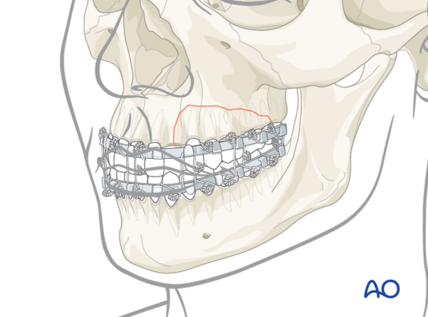
3. Exposure
In this case, the fracture is exposed through marginal incision and appropriate relaxation incisions. The fragment retains its vascular supply from the palatal side.
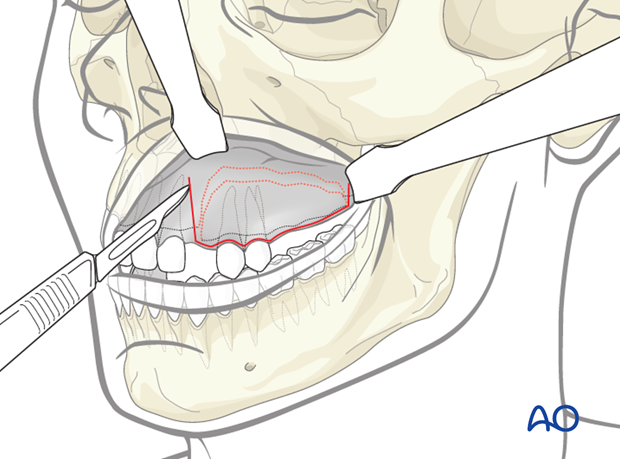
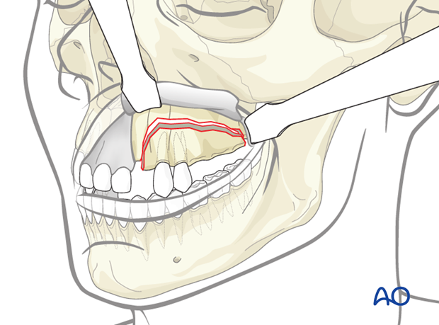
4. Reduction
Reduction is achieved by repositioning of the fractured segment under visual control. Normally, dental occlusion will lead the fragment to its correct position. This may be achieved by an assistant holding the teeth in firm occlusion. Temporary MMF with arch bars or steel ligatures may be considered.
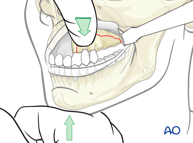
5. Fixation
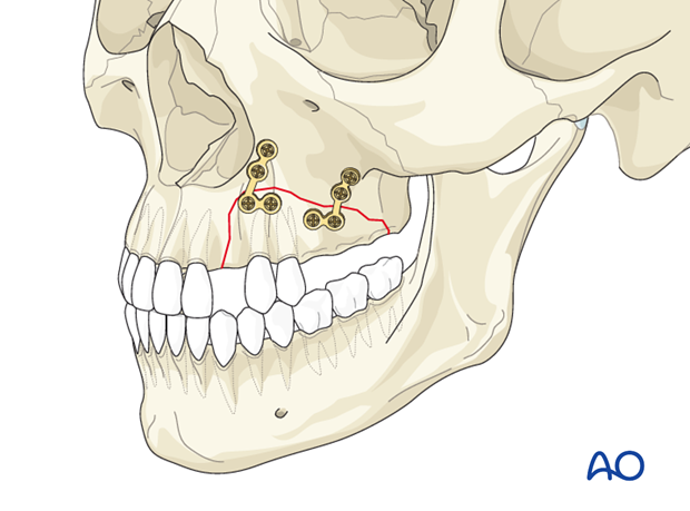
Once reduced and stable occlusion has been achieved, the fracture is stabilized with appropriate plates and screws. Care should be taken to avoid tooth roots when drilling holes for the screws. After fixation, the temporary MMF is removed and occlusion checked. Soft-tissue closure completes the surgery.
6. Aftercare following treatment of alveolar fractures
Optimal oral hygiene is essential including tooth brushing using a soft tooth brush and mouth rinsing with 0.1 % chlorhexidine twice a day. This is especially important until completion of gingival healing.
In case of undesired signs and symptoms related to the fixation appliance, the patient is encouraged to seek help as soon as possible.
Aftercare includes endodontic treatment in all teeth which develop pulp necrosis.
Follow-up
Occlusion should be regularly checked as a sign of sufficient reduction and fixation.
Diet
Patients are encouraged to restrict themselves to a semi-solid diet and avoid clenching and any other traumatic overload to the traumatized alveolar segment for a period of up to 4 weeks posttrauma.
Further treatment
Further dental treatment may be required as elective procedures, for example management of tooth fractures, discoloration, porcelain restoration, etc.


