Alveolar fracture, segmental
Definition and clinical appearance
Segmental alveolar fracture is defined as a fracture of the alveolar process which may or may not involve the socket of the teeth. The typical clinical appearance is a segment containing two or more teeth being displaced axially or laterally, usually resulting in occlusal disturbance. When mobility is tested the whole segment moves. Gingival lacerations are frequent.
Response to pulp test is almost in all cases absent. Exceptions include cases with minimal displacement.
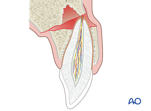
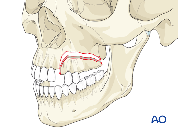
More extensive alveolar fractures
The lesion may also involve edentulous regions and present itself as a unilateral Le Fort I maxillary fracture. This requires a treatment which is different from that of tooth-bearing alveolar fractures.
The illustration shows an extensive fracture of the maxillary alveolar processes mimicking a unilateral Le Fort I maxillary fracture.
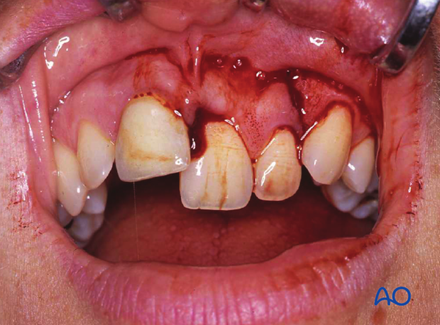
Clinical findings
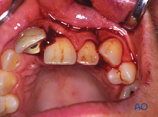
Clinical photographs show palatal displacement of an alveolar fracture comprising right central and lateral maxillary incisors.
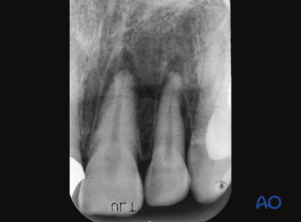
Radiographic examination
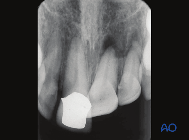
Intraoral films will show the teeth attached to the sockets and a horizontal fracture line traversing the axes of the teeth at varying levels. The fracture line mostly lies at the apical level but may present itself more distant to the tooth apices in the basal bone of the jaw. In the maxilla, the fracture may involve the maxillary sinus. The vertical fracture component usually follows the periodontal ligament of the involved teeth but it may also pass through the interdental septum as well as edentulous regions of the jaw.
Intraoral dental films, orthopantomogram, and cone beam/CT imaging are relevant image diagnostic tools.
