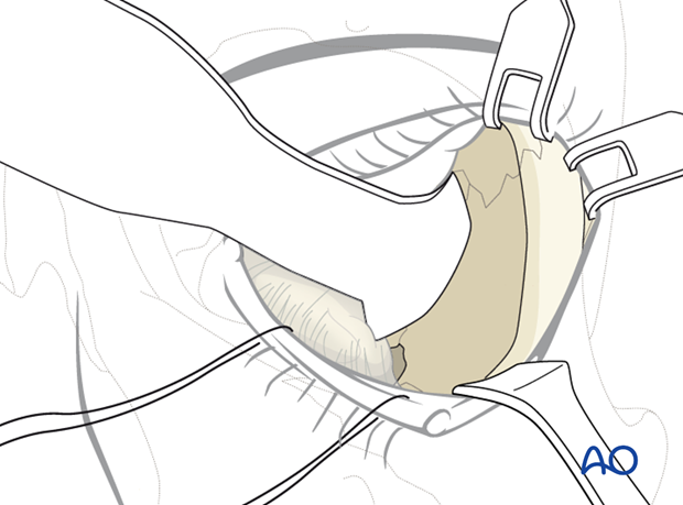
Transconjunctival approach with lateral skin extension
1. General consideration
The lower transfornix transconjunctival incision can be extended with a lateral skin incision which is then enhanced by a lateral canthotomy. The lateral extension can be combined with a transconjunctival incision via either the preseptal or retroseptal route. The approach can be started with the lateral canthotomy and continued medially into the transconjunctival approach or reversely. We describe the former option.
2. Vasoconstriction
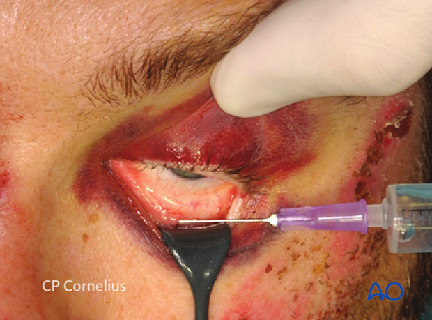
The conjunctiva in the area of the lower fornix and the area of the lateral canthotomy may be infiltrated with small amount of local anesthetic containing a vasoconstrictive agent.
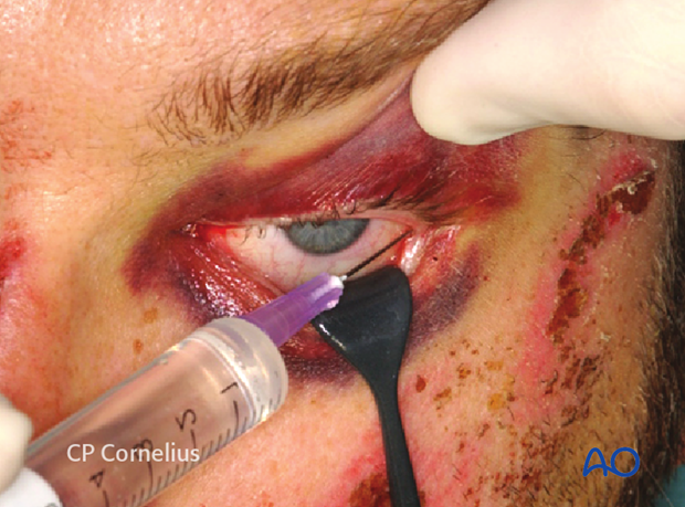
3. Corneal protection
Corneal protection as achieved with specialized shields or at a later stage of the procedure with sutures connecting the cephalic edge of the conjunctiva with the upper lid.
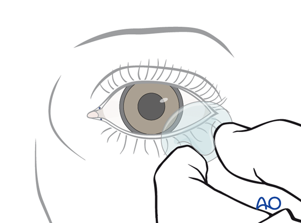
4. Lateral Canthotomy and Inferior Cantholysis
After applying traction sutures in the lower eyelid, the procedure is initiated with lateral canthotomy. A pointed scissor is inserted horizontally into the outer lid angle laterally so that the instrument contacts the underlying bone of the lateral orbital rim (approximately 7-10 mm).
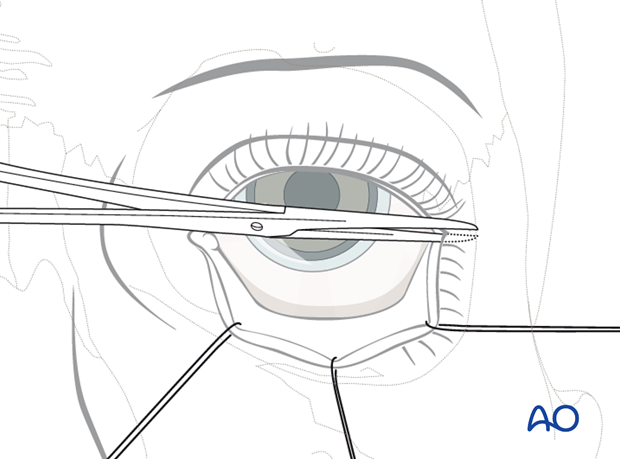
The lateral palpebral fissure is cut horizontally including the skin, the orbicularis oculi muscle and the conjunctiva. The superficial fanning fibers of the lateral canthal tendon are also transected.
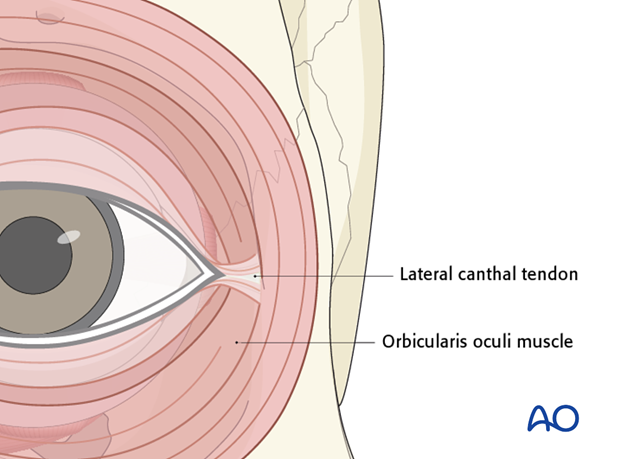
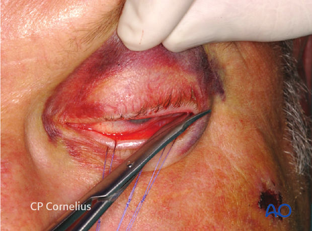
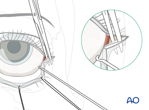
The lower eyelid is now everted using the traction sutures. It is still fixed to the lateral orbital rim by the inferior limb of the lateral canthal tendon. This part of the lateral canthal apparatus is transected (inferior cantholysis). The scissors are introduced vertically to cut the tendon. Subsequently the lower eyelid is freed and can be retracted more effectively to start swinging the lower lid outwards.
5. Transconjunctival incision
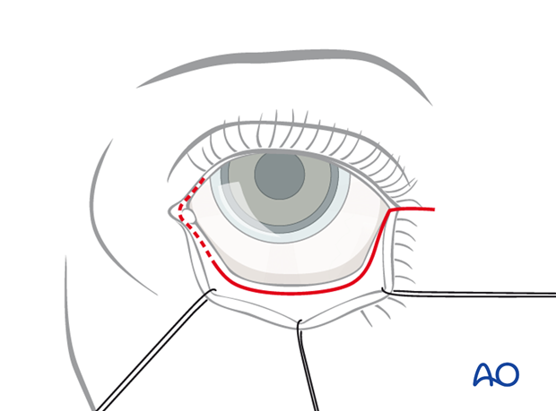
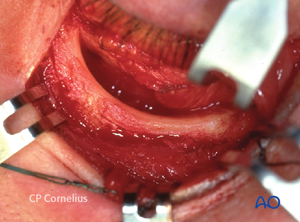
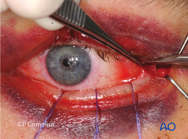
The transconjunctival incision is performed in a lateromedial direction. It can be performed preseptally or retroseptally. The lower eyelid is everted to identify the position of the lower tarsal plate through the conjunctiva.
The conjunctiva is now incised immediately along the inferior edge of the tarsus to enter the preseptal plane (for the preseptal approach, dotted line) or at the base of the fornix (for retroseptal approach, solid line).
Both techniques have been outlined previously.
The incisions are extended as far medially as necessary. With the dissection towards the infraorbital rim, the lower lid is mobilized and can be retracted anteromedially in the "swinging eyelid" fashion.
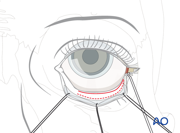
To protect the cornea against abrasion the conjunctival flap is pulled up and sutured to the upper lid margin now.
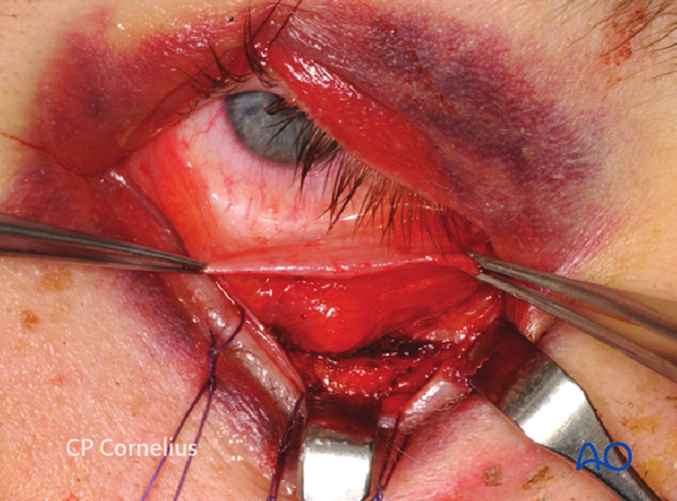
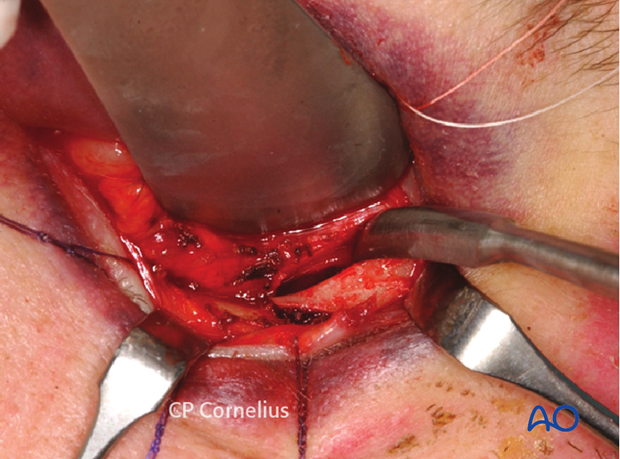
As soon as the bony surface is exposed to the desired extent, the periosteum or periorbita is incised and the dissected over the anterior maxilla or into the orbital cavity in the usual fashion.
6. Closure
The periosteum is sutured if applicable.
Prior to closing the conjunctiva, a slow-resorbing suture loop is applied exactly into the cut edges of the inferior limb of the lateral canthus and through the corresponding area of the superior limb of the lateral canthus. This loop is not yet tied, to provide open access the conjunctiva. The anchoring suture loop is tightened provisionally however to check for symmetry of the lateral canthal area to the contralateral eye and to assure that the eyelid lies adjacent to the globe.
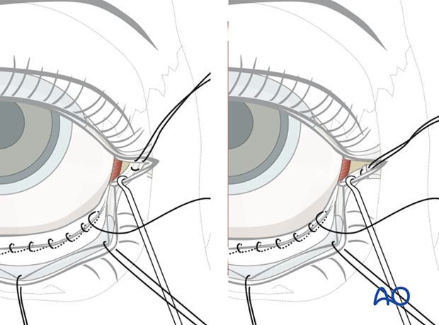
When the inferior limb of the canthal ligament cannot be identified, the a lateral tarsal strip may be exposed by elevating the skin sharply. This provide a readily identifiably stump that can be engaged to the lateral canthal apparatus.
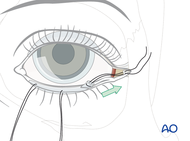
The transconjunctival incision is closed from medial to lateral while the loops for the inferior canthopexy are still loose.
The conjunctiva is usually closed with a running 6-0 fast-absorbing suture burying the knots.
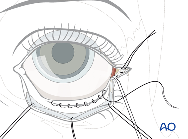
After the closure of the conjunctiva is completed, the inferior canthopexy suture is tied bringing the lower eyelid into its original position.
Subcutaneous sutures are placed along the horizontal skin incision line.
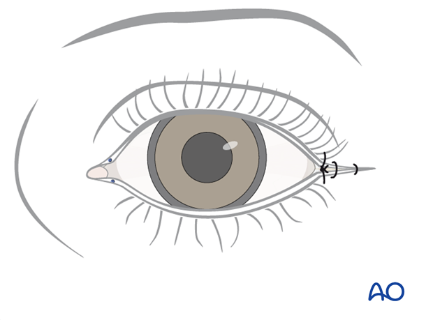
Finally, the skin is sutured with 6-0 monofilament material.
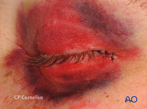
7. Superior exposure along the lateral orbital rim
The conjunctival approaches combined with the lateral canthotomy can be used to attain exposure of the entire lateral orbital rim to a level 10-12 mm above the zygomaticofrontal suture.
The exposure in cranial direction provides additional access to the lateral orbital wall.
Starting from the lateral canthotomy incision, a supraperiosteal dissection along the lateral orbital rim is performed until a point above the zygomaticofrontal suture line is reached. The orbicularis oculi muscle and the superficial fan of connective tissue fibers crossing over the lateral canthal tendon are elevated and retracted cranially during dissection.
The periosteal incision from the infraorbital rim is extended vertically upwards along the midline of the lateral orbital rim just below the retractor inserted here.
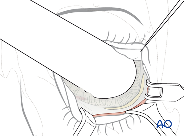
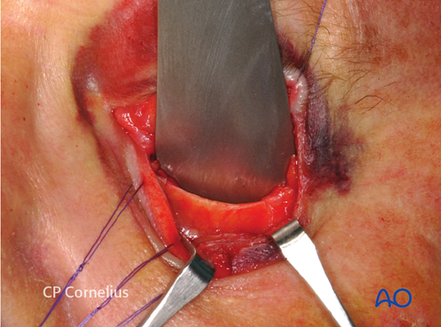
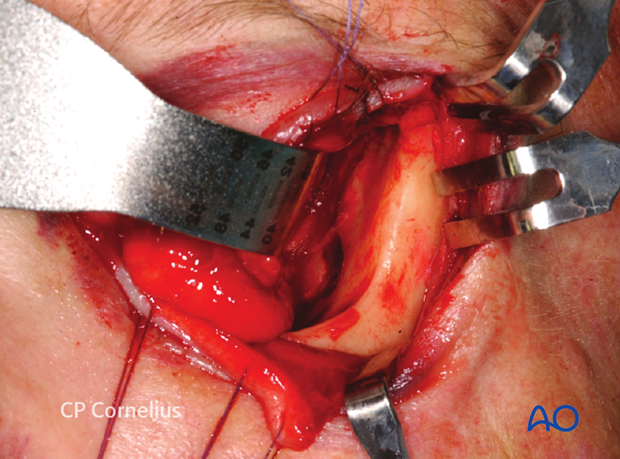
The periorbita along the inferomedial walls of the orbit is stripped from the bone including the insertion of the lateral canthal ligament at Whitnall’s tubercle.
Finally, the inferior orbital rim, the orbital floor, the lateral orbital rim, and the lateral orbital wall are exposed and the sphenozygomatic suture line becomes visible.
Closure of this approach requires refixation of the lateral canthal ligament inside the orbit and resuspension of the periorbita as well as the periosteum.
