Subtarsal approach
1. Evaluation of skin creases
This clinical photograph shows the patient of the subsequent series.

2. Marking of skin incision
Marking of the incision line is planned in a natural crease at a level below the inferior margin of the lower tarsus. If edema obscures the direction of skin creases, the contralateral eyelid determines the direction and position of the creases.
The incision is diagonally oriented and starts medially about 2–3 mm below the lid margin and courses in a laterocaudal direction.
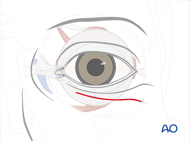
Next, the lower eye lid may be infiltrated using a local anesthetic containing a vasoconstrictor.
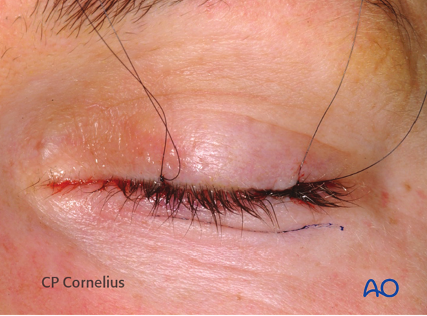
3. Skin incision
The initial incision can be made through skin and orbicularis musculature. However, it can also be performed only through the skin as demonstrated here.
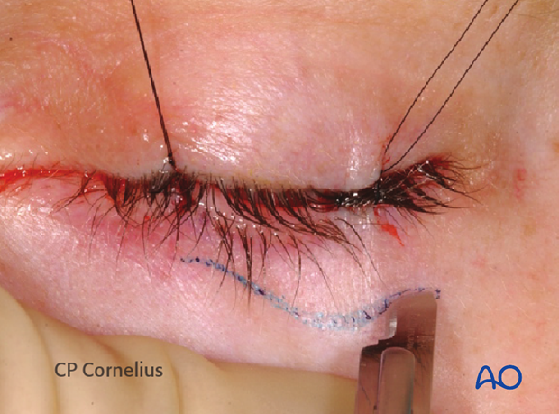
The orbicularis muscle is exposed.
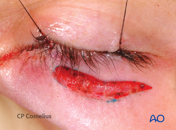
4. Dissection of the orbicularis muscle layer
The muscle is elevated laterally from the orbital septum and a small slit is opened using scissors.

Through this opening, the orbicularis muscle is undermined in the preseptal space.
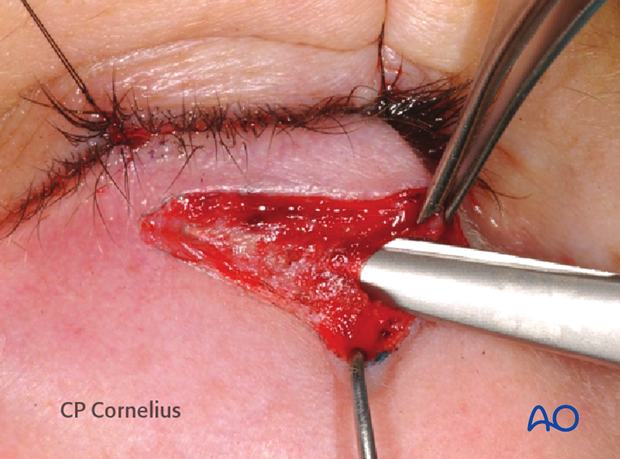
Then the muscle layer is separated from laterally to medially along the course of the muscle fibers leaving the orbital septum intact.
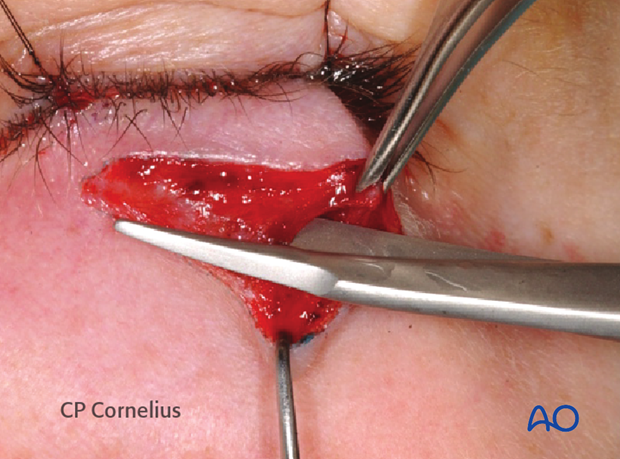
The dissection proceeds inferiorly in a preseptal suborbicular plane until the infraorbital bony margin is reached.
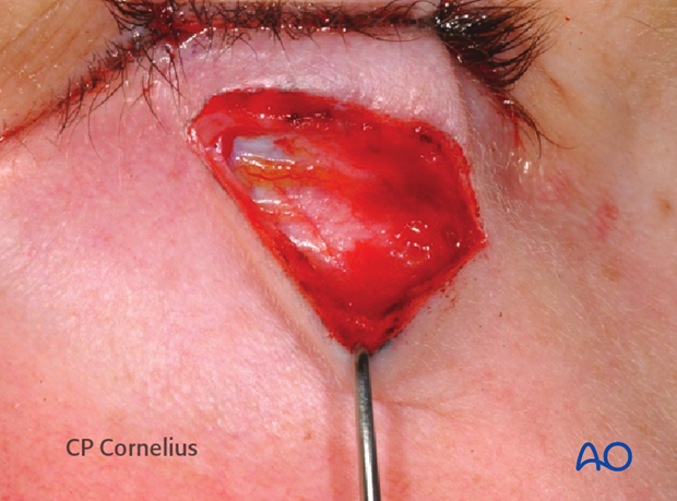
The suborbicular dissection can be carried out sharply using a scalpel, always adhering to the muscle plane.
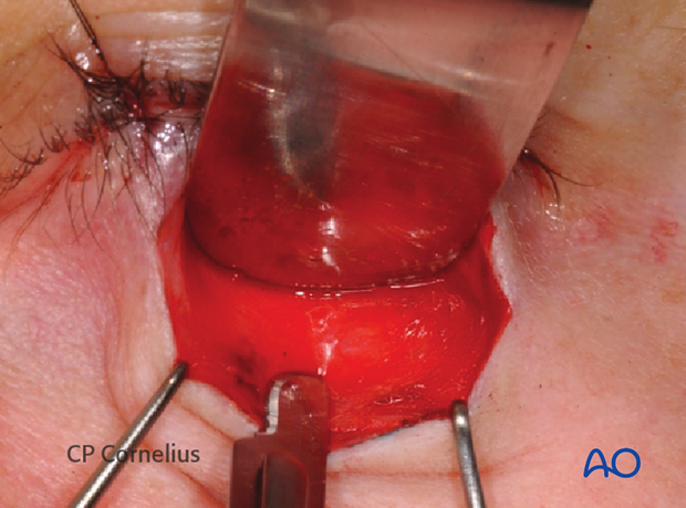
Alternatively, a pair of scissors can be used for suborbicular blunt dissection with spreading motions.
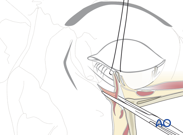
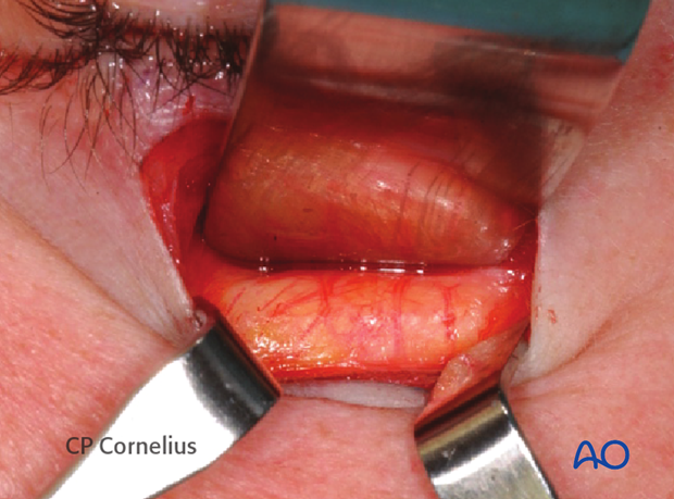
5. Exposure of infraorbital rim
The infraorbital rim is dissected in a supraperiosteal plane.
6. Subperiosteal dissection
To gain access to the orbit, the periosteum over the infraorbital rim is incised starting laterally at a level below the rim.
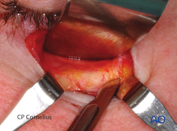
The infraorbital rim is exposed superiorly for the subsequent periorbital dissection.
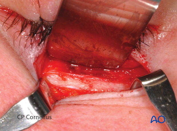
7. Periorbital dissection
The periorbital dissection is continued to expose any fractures of the orbital walls in the infero-mediolateral circumference of the bony cavity.
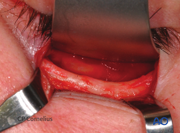
8. Wound closure
After repair of the orbital walls, the subtarsal approach is closed in layers starting with the periosteum. Next, the orbicular muscle layer is reapproximated as shown.
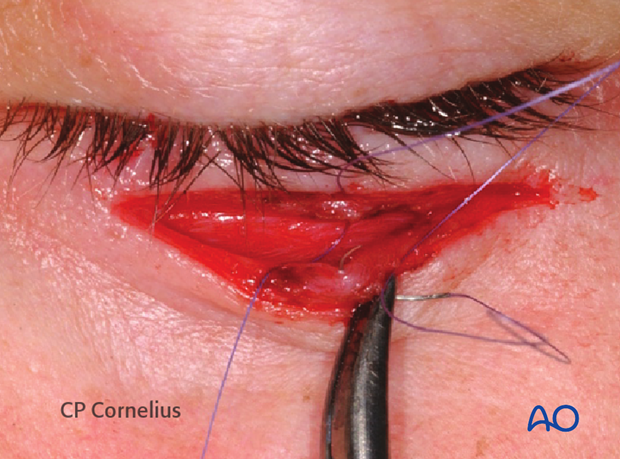
Intraoperative appearance after closure of the orbicularis muscle.
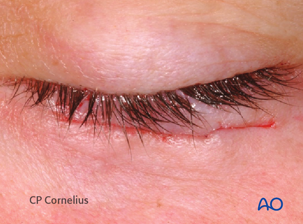
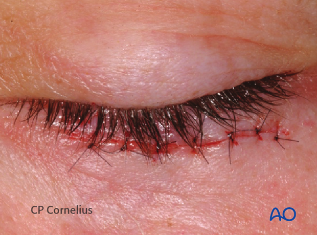
Skin closure with running or interrupted sutures. The latter is shown here.
9. Suspensory suture for lower eyelid
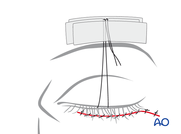
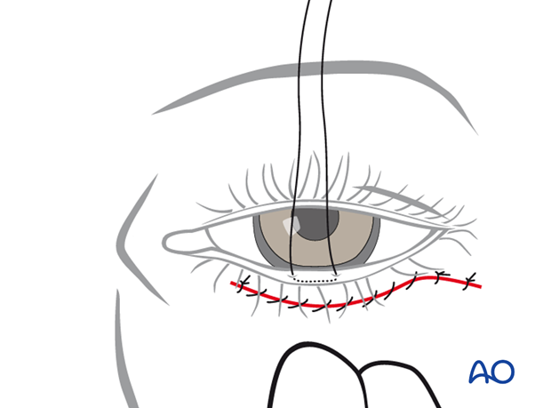
Transcutaneous lower-eyelid approaches may be complicated by vertical scar contraction during the healing period with an ectropion. Skin and septal scaring may be counteracted by so called Frost sutures.
This is a mattress suture through the Gray line of the lower lid and is applied at the end of the operation. It lengthens the lower eyelid when it is taped to the forehead. This creates traction for several days until the wound healing has passed its first critical phase.
The taping to the forehead is done in several layers in order to suspend the suture firmly and prevent downward slippage.
The suture is positioned over a first layer of tape which is applied directly to the skin. It is then secured with a second tape over it.
Next, the suture is folded over this second layer and another strip of tape secures it finally.
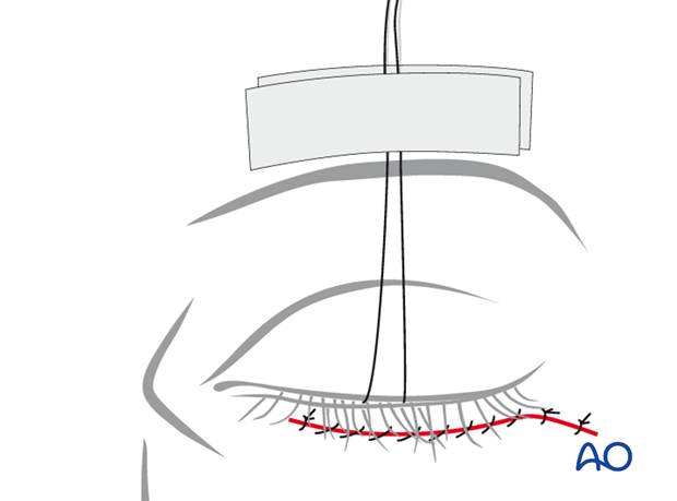
Postoperative examination of vision is possible by either removing the two uppermost layers of tape and opening both eyelids or by reflecting the upper eyelid only.
