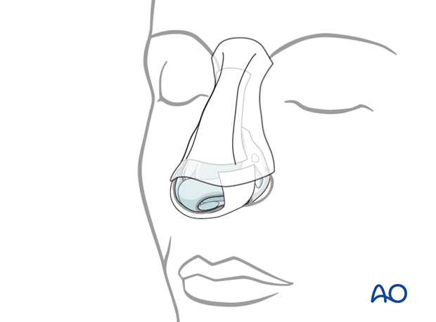
External rhinoplasty approach (open)
1. Introduction
The external rhinoplasty approach to the nasal skeleton can be used for primary treatment of traumatic injuries and for secondary procedures such as septorhinoplasty to correct posttraumatic nasal deformities.
2. Vasoconstriction and preparation
Before surgery is initiated the use of topical and injected vasoconstrictors are recommended. These should be placed at least 10 minutes prior to the incision to allow for adequate vasoconstriction.
Generally the lower nose and septum is injected with the vasoconstrictors at this time. It is important to mark the planned incision in the columella with a fine marker prior to injecting it, since the injection will distort the skin.
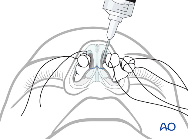
Pearl: for esthetic rhinoplasty
In some cases the surgeon is committed to the external approach from the start. In other cases a “tip delivery” approach is utilized, then the surgeon has the option of converting to an external approach, if desired.
If the tip delivery approach is selected, the incisions are made along the caudal borders of the lower lateral cartilages, but the columellar incision is not made.
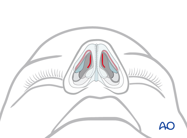
3. Incision and exposure
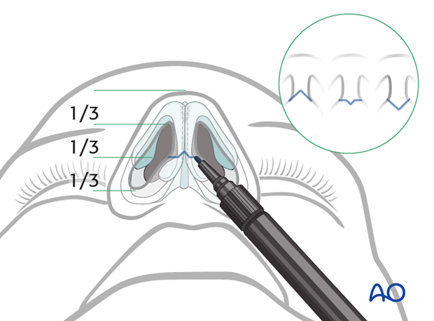
Marking the incision
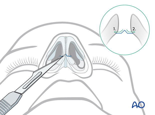
Using a fine marker, the incision of the columella is marked at approximately the junction between the proximal one third and the distal two thirds of the columella. Most people use a horizontal incision with a small “v” in the center pointed either anteriorly or posteriorly. The illustration indicates the most commonly used incisions.
Incision for the exposure of the lower lateral cartilages
The easiest way to perform this incision is to first identify the caudal border of the cartilage using the back end of the knife handle.
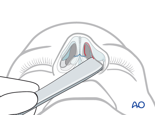
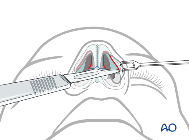
Then the mucosa is incised, taking care not to violate the cartilage itself.
The caudal end of the medial crus of the lower lateral cartilage is very close to the skin of the columella. Great care must be taken not to injure these structures when making the incision.
Exposure of the cartilages
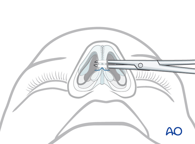
Elevation is carried out between the perichondrium and the cartilage. The perichondrium is elevated over the external surface of the lateral crus and over the medial surface of the medial crus.
Once this has been completed bilaterally, a scissor’s tip may be passed between the medial cura and the columellar skin. When the scissors are opened the columellar skin separates from the cartilages.
(The surgeon now has a choice between proceeding with a endonasal tip delivery approach or a making the columellar incision and thereby making the external approach.)
Columellar incision
Great care must be taken not to damage the columellar skin. The most precise incision is made by using the tip of a number 11 blade.
Perforate the skin at the point of the previously marked “v” and complete the incision by sawing the skin with small movements of the blade tip, taking care to maintain forward motion. Do not remove the blade from the skin until the incision is completed to one side as this may result in nick marks. Also, be careful not to violate the caudal end of the medial crus.
The knife is reinserted at the point of the “v”, and the incision is completed on the contralateral side of the columella.
Further elevation
The columellar flap is lifted with a fine skin hook (preferably a double hook to distribute the force on the skin). Elevation continues superiorly lifting the skin and pericondrium off the domes of the lower lateral cartilages, the nasal septum, and the upper lateral cartilages.
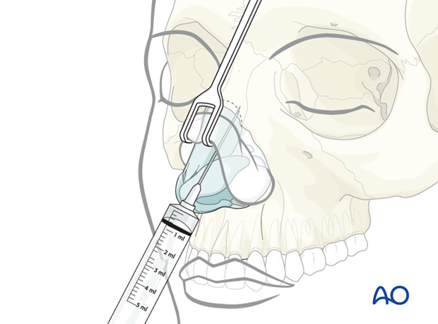
If no septal work is necessary, injection of vasoconstrictor in the area of the bony dorsum may be completed at this time.
This can be accomplished by passing the needle through the exposed area into the subcutaneous tissues overlying the bony dorsum.
Exposure of the nasal septum (if needed)
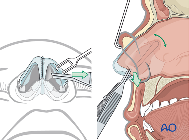
The nasal septum is located between and behind the medial crura of the lower lateral cartilages. It is exposed by separating the two medial crura.
Each medial crus is grasped with an atraumatic forceps (eg, Brown-Adson). The loose fibers between the two medial crura are divided with a scissors exposing the caudal end of the septum. Once the septum is identified, elevation of the mucoperichondrium can be accomplished on either or both sides as needed for the particular procedure.
Once the septum is exposed, the appropriate repair can be accomplished.
Exposure of the bony nasal dorsum
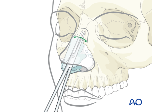
Some surgeons perform two elevations over the dorsum, while some perform one. If undraping of the dorsal nasal skin is desired, a subcutaneous elevation is carried out at this time using a blunt scissors. This frees the nasal dorsal skin, so that after the procedure it can re-drape itself into the most natural position. If a single elevation is preferred, it is accomplished in a sub-periosteal plane. An elevator or sharp scissors may be used to elevate the periosteum off the bone.
Keep in mind that if the nasal bone is fragmented, the periosteum may hold the fragments together while the repair is carried out.
4. Closure
Closure of the septum
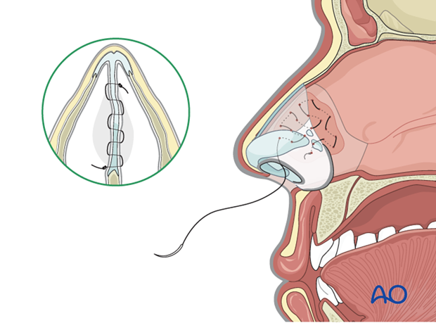
Many surgeons use a quilting suture to reapproximate mucoperichondrium to the septal cartilage to minimize the risk of postoperative septal hematoma formation. An absorbable suture is passed back and forth usually in a random fashion and then secured with a single knot. Distribute the sutures over the entire area of the septal cartilage.
Pearl: Some surgeons use a small straight needle to pass the suture back and forth. However, this often catches the mucosa on the contralateral lateral nasal wall. This can be avoided by using a curved needle.
Remaining closure
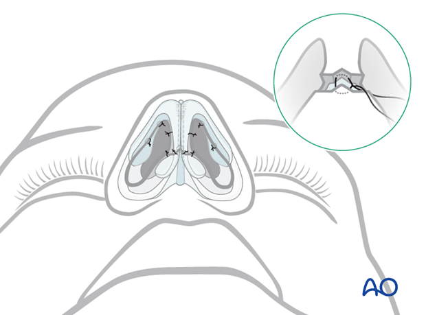
The columellar incision is closed first to ensure precise realignment. Some surgeons will first place a single deep suture, others will go directly to skin sutures typically using a 6-0 suture.
One or two absorbable sutures may be placed intranasally to reapproximate the mucosal edges. Take great care to ensure that the lower lateral cartilages are not distorted by these sutures. This is best accomplished by grasping only the very minimum of mucosa with each stitch.
Nasal packing may be placed for hemostasis and support according to the surgeon’s preference and particular circumstances.
External dressing
A variety of external dressings can be applied. Most surgeons tape the nasal dorsum after applying an adhesive to the skin. A rigid splint of some type is commonly applied over the tape. Care should be taken to avoid wrinkles in the tape which may form pressure points under the splint.
