Anatomy of the facial nerve
1. Anatomy of the facial nerve
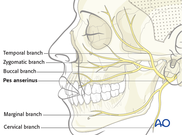
The facial nerve courses through the temporal bone and exits the stylomastoid foramen. The location and position of the facial nerve at the stylomastoid foramen is consistent. The main trunk branches at the pes anserinus into the upper and lower divisions.
The traditional description includes five peripheral main branches of the facial nerve. These are: temporal, zygomatic, buccal, marginal, cervical branches. However, there is considerable variability in the branching pattern beyond the pes anserinus.

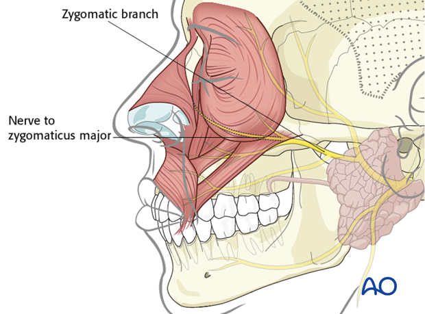
The zygomatic branch is the most important for eye closure and commissure elevation (smile). There is a branch arising from the zygomatic that supplies both the zygomaticus major muscle proximally and the palpebral portion of the orbicularis oculi distally. This nerve is referred to as the nerve to zygomaticus major. It innervates the palpebral portion of the muscle and is the primary source of blinking and eye closure. Because of the dual innervation this nerve provides, it is extremely difficult to completely separate the function of the zygomaticus major (smile) from the orbicularis oculi (blinking). This is the most common cause of synkinesis.

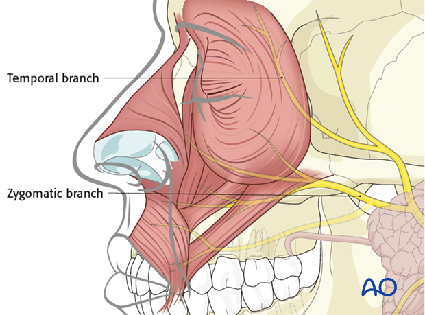
The orbicularis oculi (orbital portion) is also supplied by branches from the temporal and zygomatic nerves. This innervation is mainly responsible for voluntary eye closure and not spontaneous blink. However, during reinnervation these branches can be used to improve tone of the orbicularis oculi muscle and eye closure.

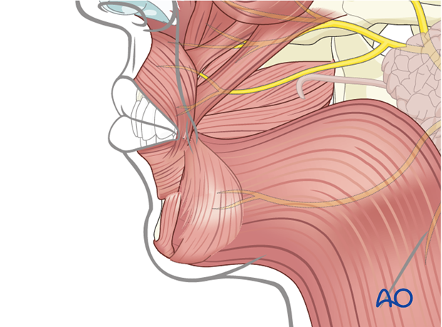
The lower division of the facial nerve is not responsible for the smile (commissure elevation). It is primarily responsible for lower lip depression, pucker, and platysma function.


