Scapula
1. Introduction
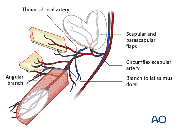
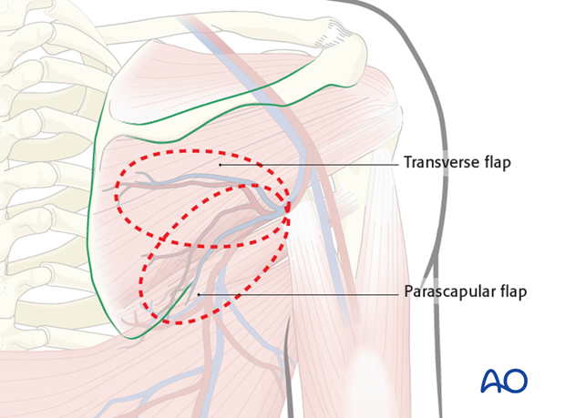
The scapular system provides the option between the following flaps
Fasciocutaneous flap
- Transverse Scapular flap
- Parascapular flap
Myocutaneous flap
- Latissimus dorsi +/- anterior serratus muscle flap
Osteomyocutaneous flaps
- Scapular bone flap +/- parascapular flap
We will here describe their individual harvest. However, all the above flaps can be harvested together as a chimeric flap.
Anatomy
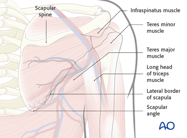
The surgical landmarks of the scapula are as follows
- The lateral border of the scapula
- The scapular spine
- The scapular angle
- The triangular space created by the three muscles
- Teres major muscle
- Teres minor muscle
- Long head of triceps muscle
- The latissimus dorsi and anterior serratus muscle
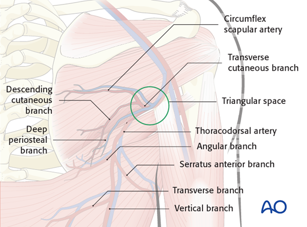
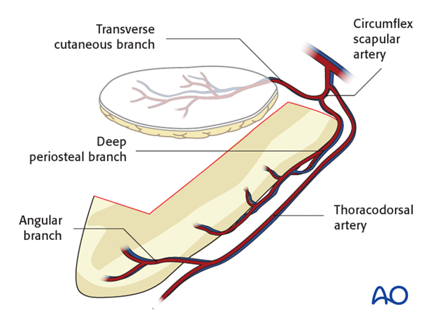
The main trunk of the scapular vascular system is the subscapular artery. This artery is the origin of two main branches, the circumflex scapular and the thoracodorsal artery.
The Circumflex scapular divides into the following branches of relevance:
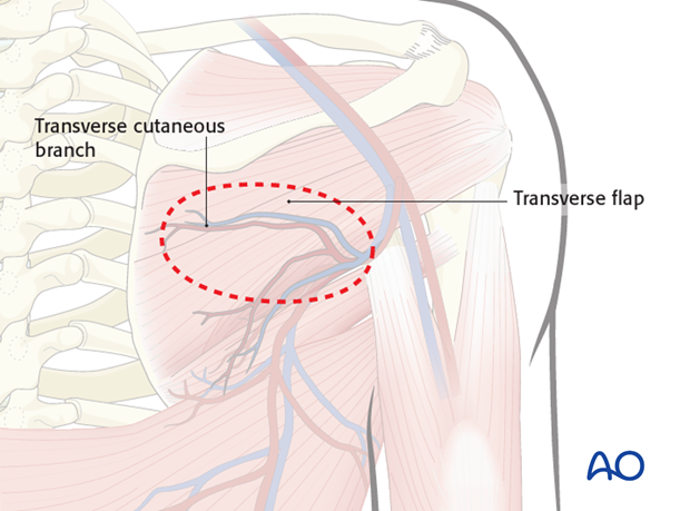
- Transverse cutaneous which supplies the scapular flap
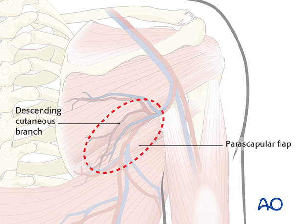
- descending cutaneous which supplies the parascapular flap
- The deep periosteal branch which supplies the scapular bone flap.
The thoracodorsal artery divides into the following branches of relevance:
- The angular branch, vein supplies the scapular angle
- The branch to the serratus anterior which supplies the serratus anterior muscle flap
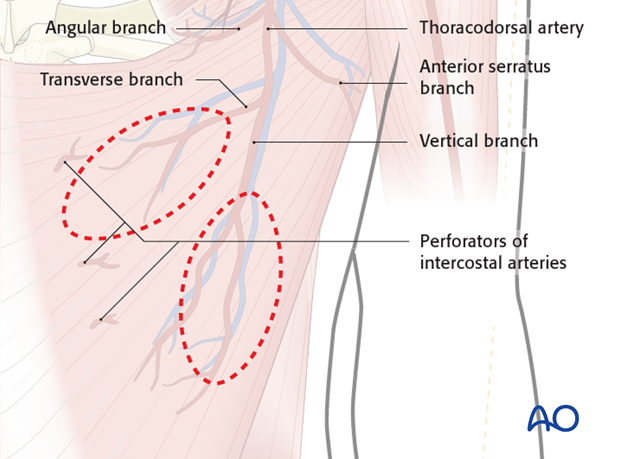
- The transverse and vertical branch to the latissimus dorsi muscle
2. Transverse/para scapular flap
Flap blood supply
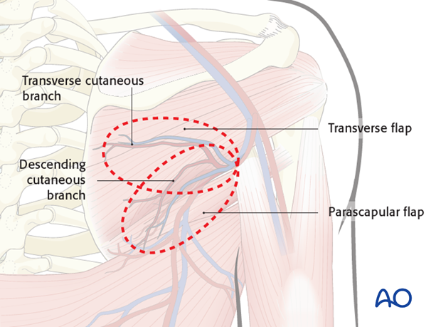
The transverse scapular flap (transverse flap) receives its blood supply from the transverse branch of CSA & venae comitantes.
The parascapular flap receives its blood supply from the descending branch of CSA & venae comitantes.
Flap design
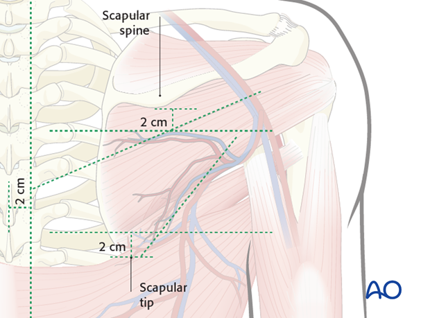
The flap is harvested inferior to and in parallel with the scapular spine. The skin paddle has to incorporate the triangular space.
The limits of the skin paddle design is:
- 2cm below the scapular spine,
- 2cm above the scapular tip,
- 2cm lateral to the midline
- Maximum length: 24 cm
- Superior to the lateral scapular border
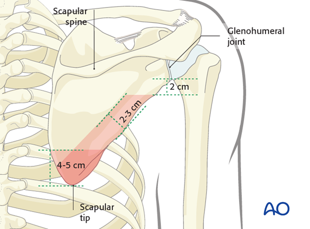
The bone flap is limited in size by the following borders:
- 2-3 cm medially from the lateral border of the scapula
- 2 cm inferiorly to the glenohumeral joint
- If only the scapular tip is harvested, maximum 4-5 cm is available measured from the tip.
Flap harvest
The anatomical landmarks are marked and the skin flap outlined within its maximum limits.
If a smaller flap is indicated, it is advisable to perform a pre-operative ultrasound Doppler to identify the location and path of pedicle.
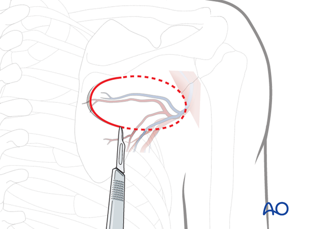
To illustrate the procedure, we will show the harvest of the transverse flap. The harvest of the parascapular flap follows the same principle.
Starting medially, the skin is incised down to the deep fascia of the skin and a subfascial elevation from the infraspinatus muscle is performed.
As the flap is raised, the pedicle on the undersurface of the cutaneous layer can be identified.
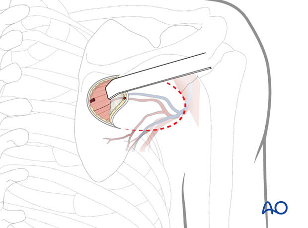
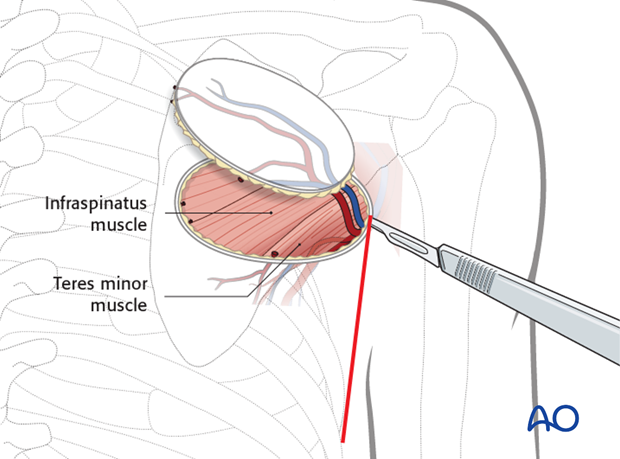
A vertical skin incision is made at the lateral edge to allow for the retraction of the skin and the dissection of the pedicle.
The dissection is continued laterally until the teres minor muscle is encountered and the origin of the pedicle can be found in the triangular space.
Meticulous dissection is performed expose the circumflex scapular artery up to the bifurcation (maximum limit) of the subscapular artery.
The skin incision is completed and the flap is mobilized from the underlying muscles.
When scapular bone is not needed, the cutaneous flap is now ready and can be transected when the recipient site is ready.
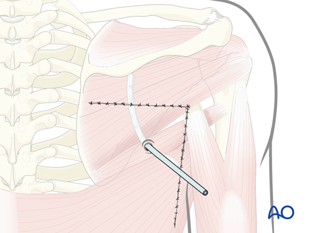
A drain is inserted and a primary wound closure performed.
Bone harvest
While taking great care not to damage the CSA, dissection along the CSA is performed until the subscapularis artery is encountered.
If bone is required, The deep periosteal branch of the CSA can be identified as running inferiorly and entering the lateral border of the scapula.
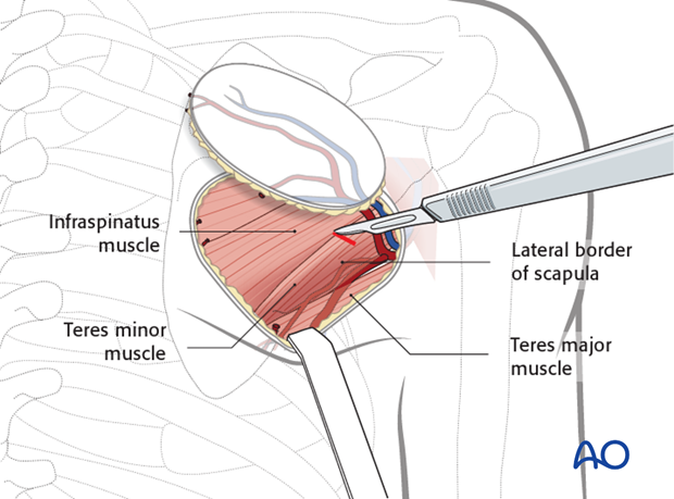
The teres minor is incised 3 cm medially from the lateral border of the scapula and retracted medially to expose the bone. A small cuff of the teres minor is left attached to the scapular bone in order to protect the blood supply of the bone flap.
The teres major and part of the latissimus dorsi muscles which are attached to the segment that is harvested are transected.
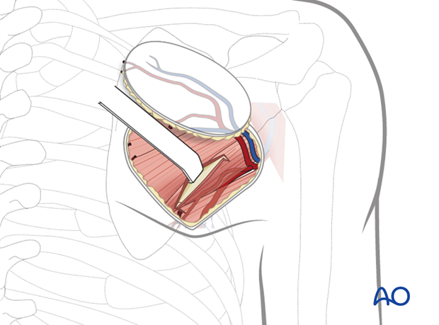
The teres major is retracted superiorly and an osteotomy is performed as planned with a saw.
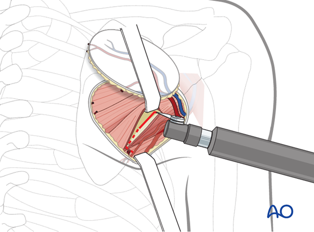
The bone is retracted laterally and freed from the subscapularis muscle (deep part of scapular bone).
Taking care not to compromise the CSA and the deep periosteal branch, the teres minor still attached to the bone is transected.
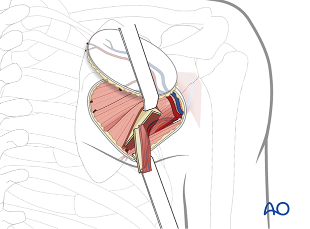
The flap is now completely mobilized and the pedicle is transected when the recipient site is ready.
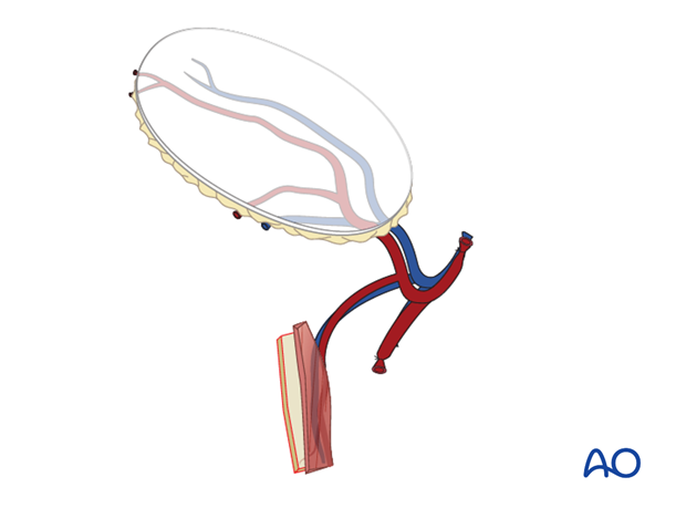
The teres major is repositioned, a drain is inserted and a primary wound closure of the skin is performed.
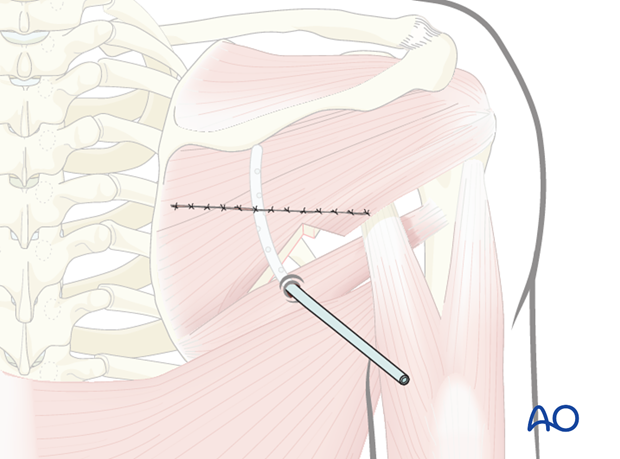
3. Latissimus dorsi
Flap design
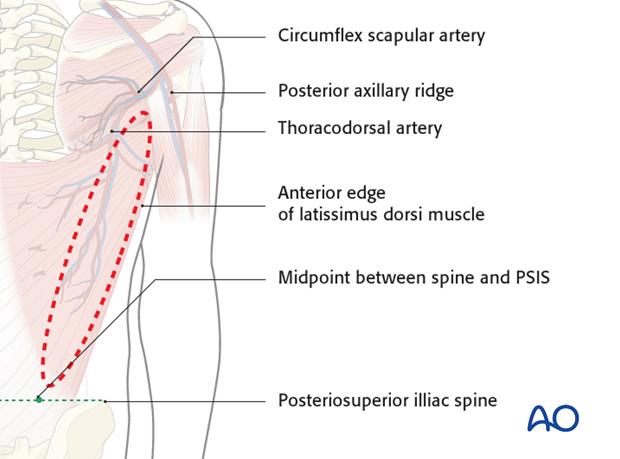
The latissimus dorsi flap receives its blood supply from the thoracodorsal artery.
The essential landmarks are:
- Posterior axillary ridge
- Anterior edge of the latissimus dorsi muscle
- Posteriosuperior iliac spine (PSIS)
- Midpoint of imaginary line between PSIS and thoracolumbar spine
The flap can be harvested with an axis parallel to the muscle fibers.
The lateral edge of the skin paddle should not be designed beyond anterior edge of the latissimus dorsi.
The flap can be harvested to a length of 21-38 cm and a width of 7-14 cm.
As the skin receives it blood supply from the latissimus dorsi, several skin paddles can be prepared.
If needed two separate myocutaneous paddles may be prepared based on the transverse and vertical branch of the latissimus dorsi.
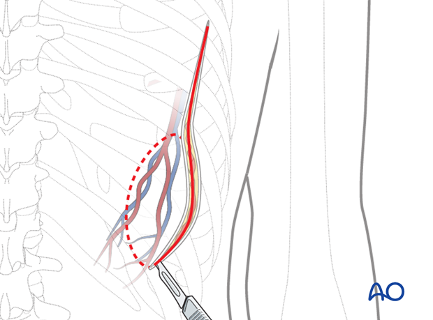
The anatomical landmarks are marked and the skin flap outlined within its maximum limits.
The inferior incision is made to the deep fascia of the skin and the anterior edge of the latissimus dorsi is identified.
For obese patient, it is advisable to pass a few anchorage sutures to secure skin to muscle, prevent torsion over the tiny skin perforators.
The latissimus dorsi is retracted medially and the pedicle (located 2cm medially to the anterior edge of the latissimus dorsi) is dissected and protected.
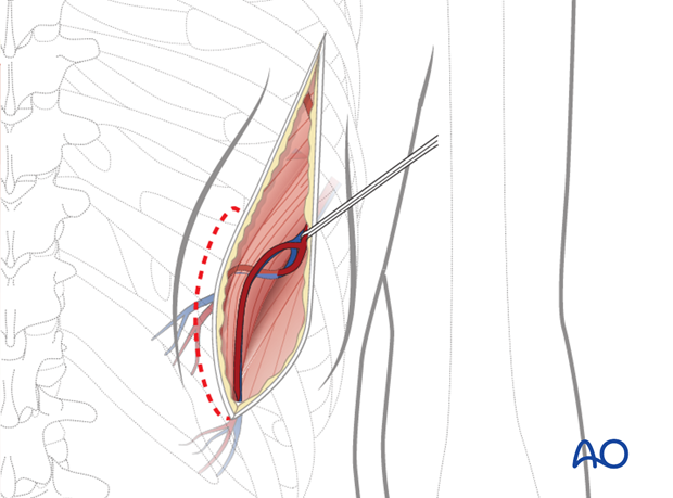
The skin incision can be completed.
The thoraco dorsal pedicles are running beneath the latissimus dorsi muscle, entering the hilum as it courses caudally.
The anterior serratus branch running anteriorly into the serratus muscle is ligated and transected if anterior serratus flap is not required. Right opposite to it, angular branch is transected and ligated.
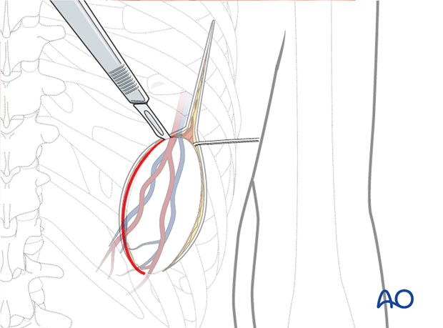
The latissimus dorsi is transected medially as needed to obtain the correct size of the flap.
Meticulous dissection is performed along the thoracodorsal artery up to the teres major muscle.
The teres major muscle is retracted and the dissection continued until the bifurcation of the subscapular artery is reached.
When the recipient site is ready, the thoracodorsal pedicle is transected.
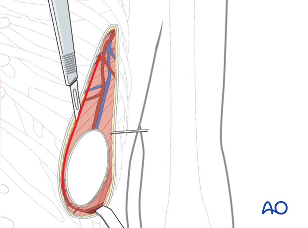
A drain is inserted and a primary wound closure performed.
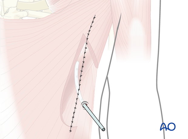
If a chimeric flaps is harvested (cutaneous scapular flaps, scapular bone flaps and the latissimus dorsi flaps) . The teres major muscle will also need to be transected. Now, the subscapular artery is ready to be transected and harvested along with the chimeric flaps.
