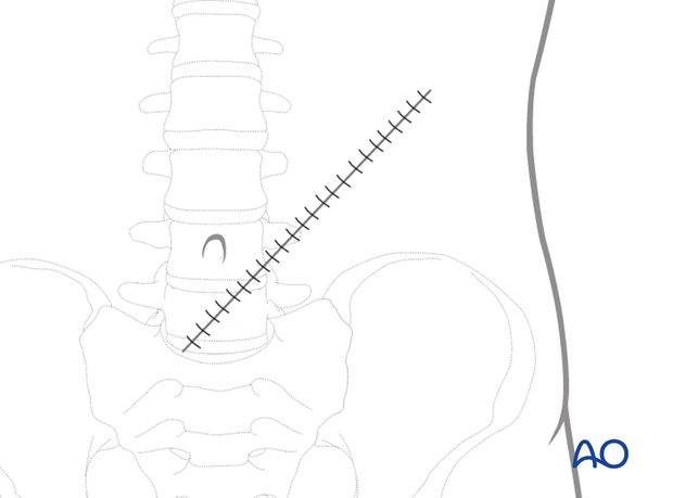
Rectus abdominis
1. Introduction
Patient positioning
The rectus abdominis flap can be transferred as a muscle or myocutaneous free flap.
Perioperative assessment must include any history of prior abdominal surgery to ensure the integrity of vascular pedicle.
Anatomy
The rectus abdominis muscles are located in the paramedian region of the anterior abdominal wall.
Each muscle originates from the pubis and inserts into 5th, 6th and 7th ribs and the xyphoid process.
The following landmarks are identified:
- Costal margin
- Linea alba
- Linea semilunaris
- Iliac crest
- Pubis
- External iliac vessels
- Femoral artery (palpation for pulse)
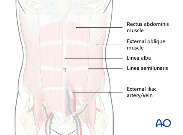
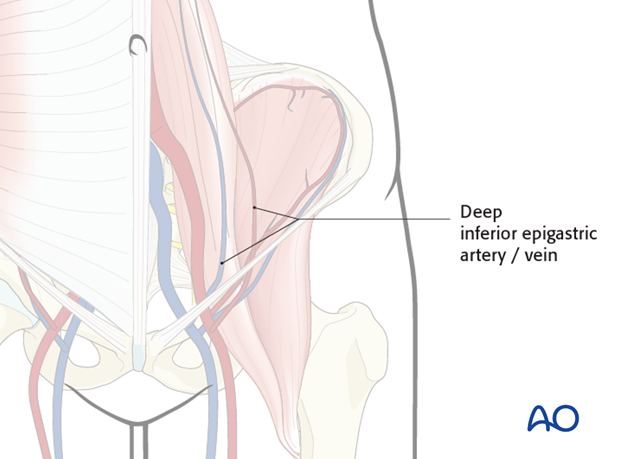
The vascular supply is the deep inferior epigastric artery and the deep inferior epigastric vein.
Flap design
There is a versatility in the design of the skin paddle based on the perforators located in the periumbilical region.
Transverse skin paddles can be designed:
- Horizontally
- Vertically
- Transversely
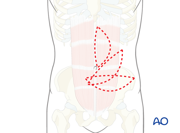
When a muscle flap is harvested, a portion or the entire rectus abdominis muscle can be harvested based on the deep inferior epigastric system.
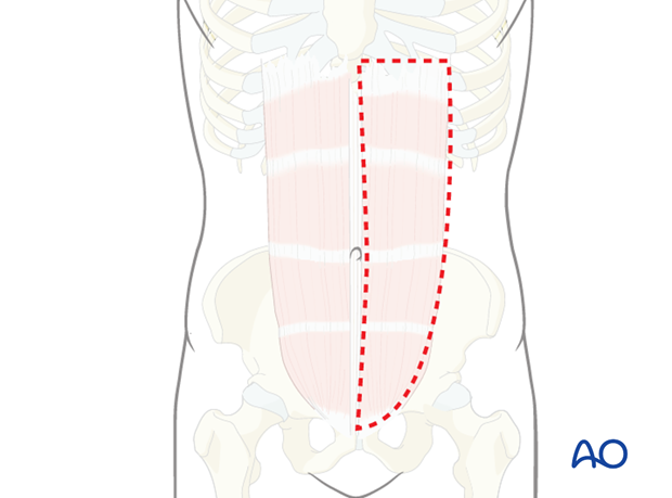
The outline of the rectus abdominis muscle can be drawn on the anterior abdominal wall prior to designing the skin paddle. The femoral artery is palpated and drawn as a landmark together with the outline of the linea semilunaris.
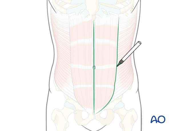
A transverse skin paddle is outlined in the periumbilical region centered on the course of the rectus abdominis muscle.
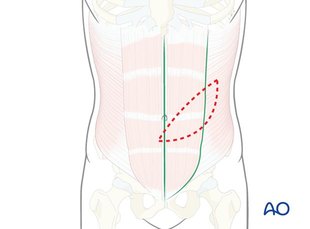
Skin incision
The skin incision is made at the superior border of outlined paddle through the subcutaneous tissues to the level of the fascia.
The rectus abdominis is identified after incising the entire rectus sheath from the linea alba to the linea semilunaris.
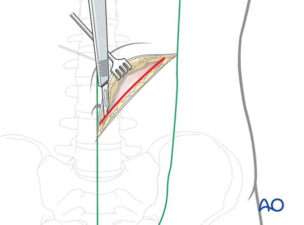
The inferior skin incision is then completed as described for the superior incision.
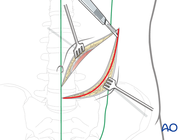
The lateral portion of the skin paddle is elevated off the external oblique muscle to linea semilunaris.
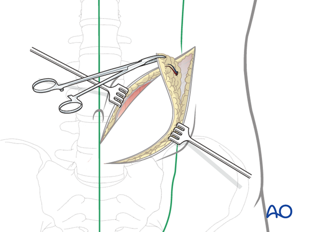
The musculocutaneous perforators are now identified and protected.
The anterior sheath is incised laterally, medial to the linea semilunaris preserving the perforators.
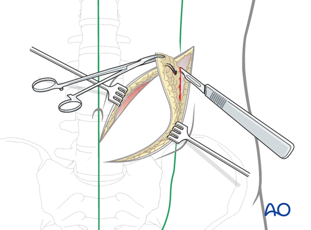
The medial portion of the skin paddle is elevated off the contralateral rectus sheath.
The musculocutaneous perforators are now identified and protected.
The anterior sheath is incised lateral to the linea alba preserving the perforators to the skin.
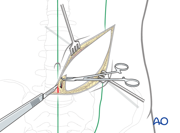
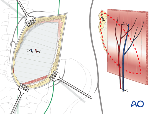
The rectus abdominis muscle is transected above the level of the skin paddle and the muscle elevated off the posterior rectus sheath using blunt dissection.
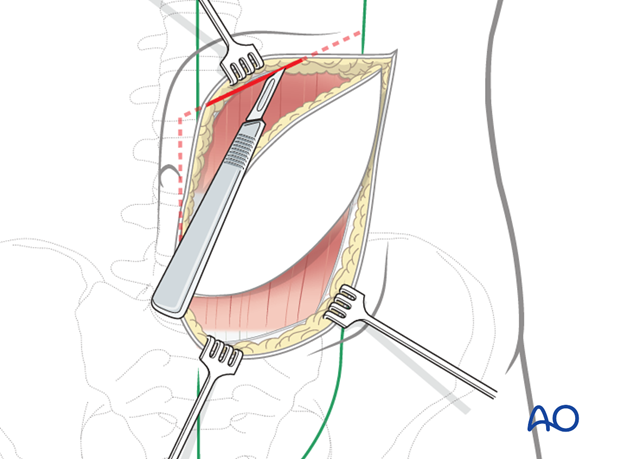
As the dissection proceeds inferiorly the deep inferior epigastric pedicle is identified.
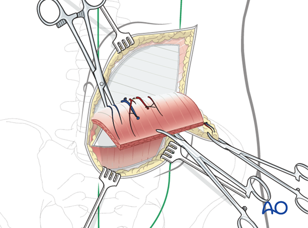
The dissection of the pedicle is continued to the level of its takeoff from the external iliac artery and vein.
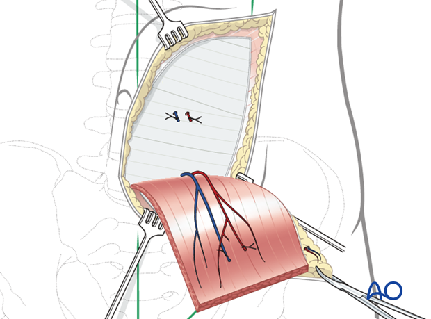
Depending on the amount of muscle required for the reconstruction, the inferior transection of the rectus muscle is performed, protecting the vascular pedicle.
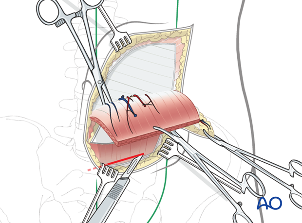
When the recipient site is ready, the pedicle is transected.
2. Closure
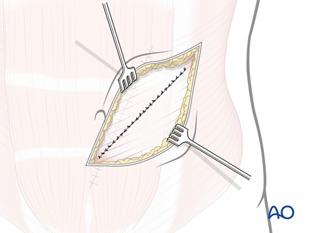
Meticulous closure of the donor site must be performed to prevent an abdominal hernia.
The anterior rectus sheath must be repaired to the level of the arcuate line.
The preserved cuffs of the anterior sheath above the arcuate line are reapproximated when possible to reinforce the abdominal wall closure.
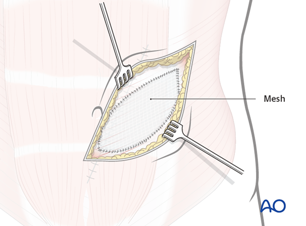
Alternatively a mesh can be sutured from the linea alba to the linea semilunaris above the arcuate line to reconstruct the anterior abdominal wall.
Closure of the skin is accomplished by undermining both laterally and medially.
