Radial forearm fasciocutaneous flap
1. Introduction
Anatomy
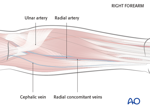
The blood supply of the flap is based on the radial artery, radial concomitant veins (deep system) and the cephalic vein (superficial system).
The following landmarks should be identified and marked prior to surgery:
- The radial artery (identified by palpation)
- The cephalic vein
Flap design
The flap can be harvested in a size up to 8 x 16 cm.
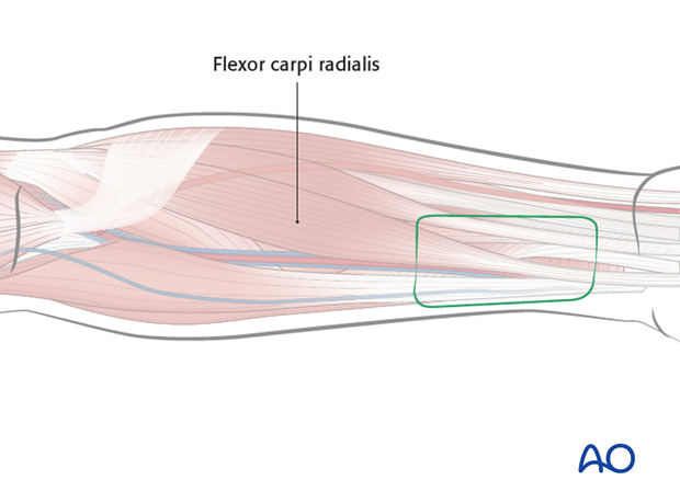
The flap should be centered between the cephalic vein (if this vein is to be used) and the radial artery. If cephalic is not reliable or if radial concomitant vein is selected, the flap will be centered at radial artery.
The palmar border of the flap should be placed
over the flexor carpi radialis.
2. Flap harvest
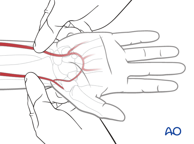
Allen's test
An Allen's test is performed to ensure the integrity of the palmar circulation prior to the harvest of the radial forearm flap.
Placement of tourniquet
A tourniquet is placed on the upper arm and inflated to a pressure of 1.5x patient’s systolic pressure (usually 250 mmHg). The tourniquet should not be applied for more than 60 min.
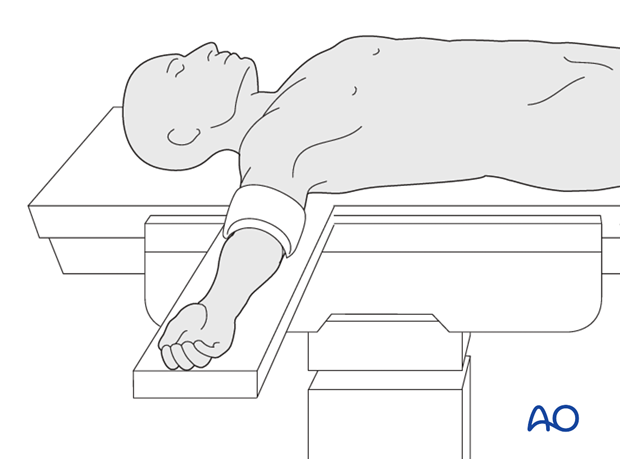
Flap outline
The flap outline is marked on the skin together with a proximal lazy S extension of the incision toward the antebrachial fossa.
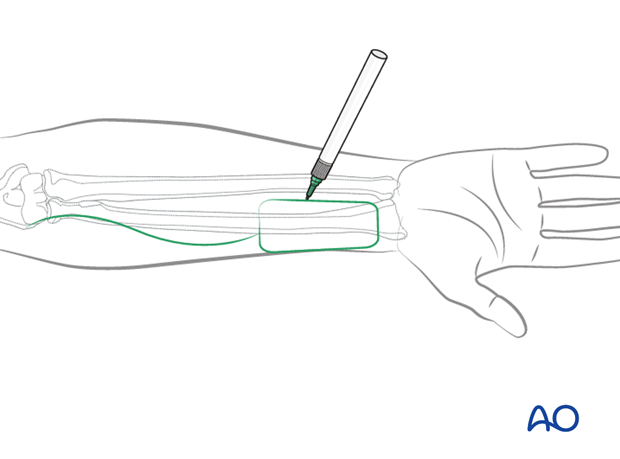
Skin incision
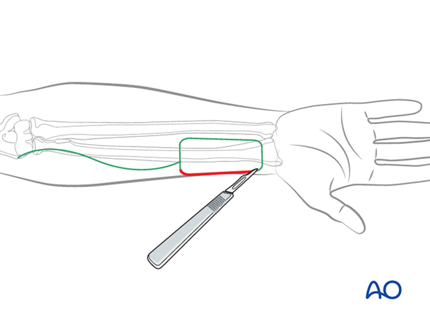
The skin incision is performed as outlined.
Lateral incisions can be carried down to the deep fascia of the skin taking care to keep underlying paratenon intact.
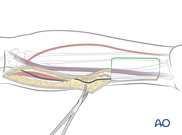
Mobilization of cephalic vein
The wavy skin incision is made down to the subcutaneous tissues and raised laterally, the cephalic vein is identified. Starting distally, the cephalic vein is mobilized together with the surrounding subcutaneous adipose tissue.
Ligation of distal vessels
Blunt dissection for the cephalic vein, radial artery, concomitant veins and the lateral antebrachial cutaneous nerve is performed at the distal end of the flap.
The distal end of the cephalic vein, radial artery and radial vein are transected and ligated.
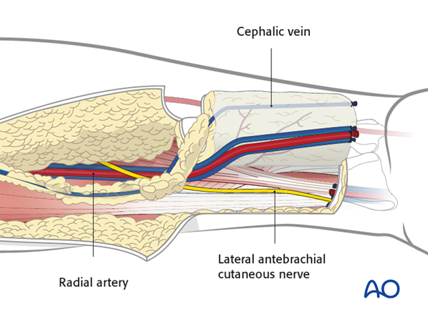
In case the sensate flap is harvested, lateral antebrachial cutaneous nerve branches are also transected at this point.
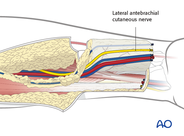
The distal dissection can then be carried down to the deep fascia. On the radial side of the flap, care should be taken to preserve the superficial radial nerve branches to the wrist. The flap can then be raised together with the vascular bundle of the flap (deep system consisting of the radial artery and vena comitantes) and the cephalic vein from distal to proximal (superficial system).
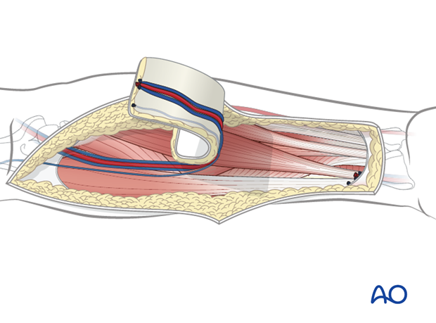
Mobilization of vascular bundle
The brachioradialis is retracted laterally and the vascular bundle located in the groove between the brachioradialis and flexor carpi radialis muscles is mobilized taking care that all its branches (perforators to the muscles) are ligated.
The pedicle dissection along the intermuscular septum is continued to the level of the brachial artery. The flap is then re-perfused by releasing the tourniquet to verify the viability of the fasciocutaneous flap.
In addition it is important to also assess the vascularity of the hand at this point.
Hemostasis is obtained and the vascular pedicle is then divided when the recipient site is ready.
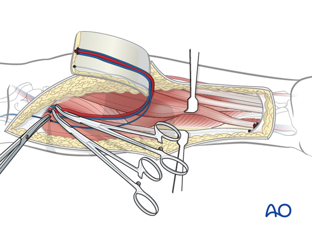
Closure
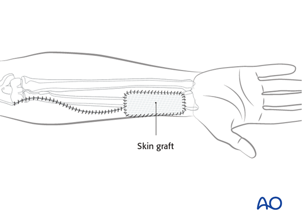
If the skin cannot be closed by primary closure, a skin graft is harvested, trimmed to cover the skin defect of the donor site and sutured in place. The graft is covered by a compression packing for 10 days.
