Pectoralis myocutaneous pedicle flap
1. Introduction
Anatomy
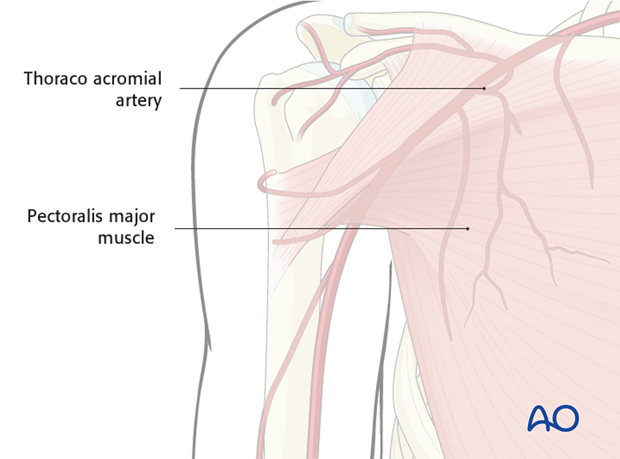
The flap receives its blood supply from the thoraco acromial artery and the secondary segmental perforators arising from the internal mammory artery.
The lateral thoracic artery does not usually contribute significantly to the vasculature of the pectoralis muscle.
Flap design
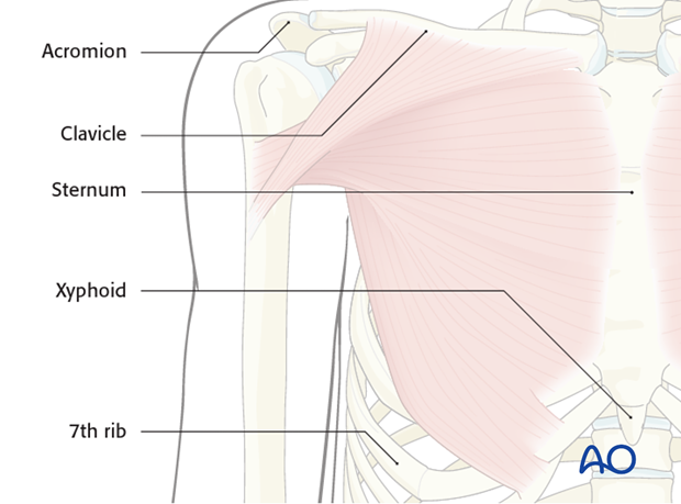
The following landmarks are identified and marked:
- Acromion
- Clavicle
- Sternum
- Xyphoid
- 7 th rib
Size limitations
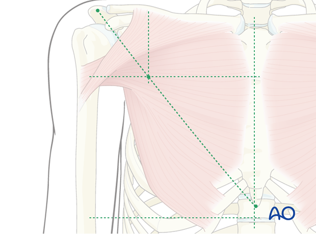
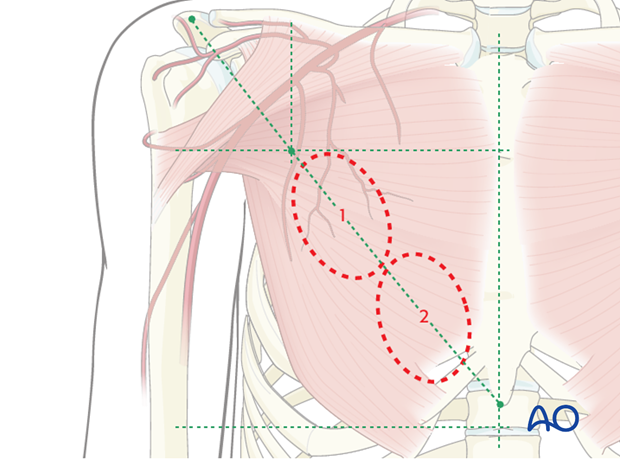
A line from the acromium to the tip of the xyphoid is drawn. Then a second line is made perpendicular to the clavicle starting between mid-clavicle and the distal third of the clavicle. The crossing point of the two lines is used as the superior limit of the skin paddle.
The limits of the skin paddle are:
- The 7 th rib inferiorly.
- Laterally the extent of the pectoralis major muscle.
- Medially the border is at the midline of the sternum
- The skin paddle position over the muscle will depend on the location of the neck/oral defect. The skin paddle illustrated in this procedure is suitable for a neck defect. For oral defects the skin paddle would be positioned more inferomedially.
The pectoralis major muscle is harvested in full thickness. The width and length of the muscle harvested will depend on the size of the defect.
The skin flap is designed as a curved ellipse to facilitate wound closure.
Here the skin paddle is outlined fairly proximally (1) limiting the arc of rotation of the flap, making it suitable for neck defects.
Should the surgeon need to reconstruct an intraoral defect, or cheek defect, placing the skin paddle more distally (2) allows for a greater arc of rotation and hence more reach of the skin paddle.
2. Flap harvest
Exposure of the pectoralis major muscle
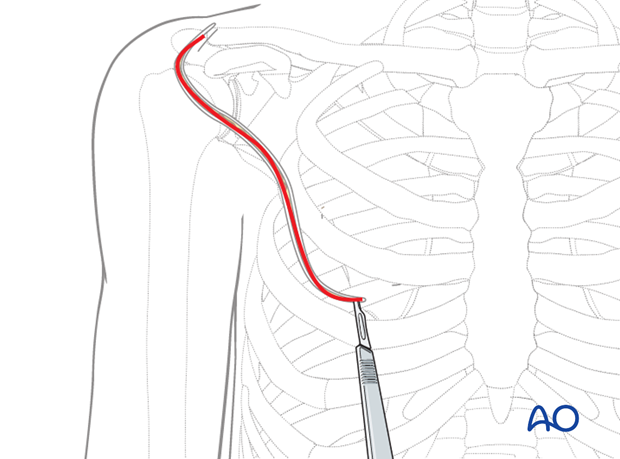
The lateral skin incision is made down to the deep fascia and raised laterally to expose the pectoralis major muscle.
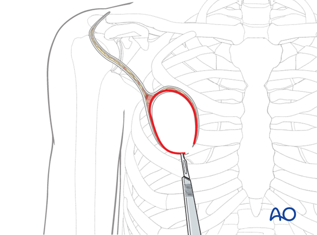
The medial skin incision is made down to the deep fascia and raised medially to expose the pectoralis major muscle.
Part of the rectus sheath can be seen running longitudinally at inferior edge.
Raising of the muscle
Depending on the location of the skin paddle, the rectus sheath is incised and raised superiorly together with the pectoral major muscle from the external intercostal muscles and ribs.
The internal mammary perforators are transected and ligated.
Digital dissection can be performed after rib nr. 4.
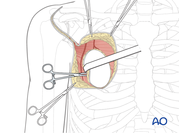
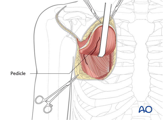
Upon raising the PMMF, pedicle can be seen running beneath the pectoralis major muscle and entering the muscle hilum at the location medially to pectoralis minor muscle. Pedicle can be felt via pulse palpation.
Dissection of the pedicle
The thoraco acromial pedicle can be identified medially to the pectoralis minor muscle.
The lateral thoracic artery is ligated as the insertion of the pectoralis major muscle to the humeral head is transected laterally. However, in some cases where the lateral thoracic is the dominant vessel, then the lateral thoracic artery should be preserved as it is the main blood supply to the muscle.
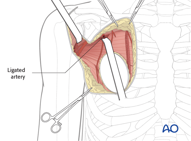
The three main branches of the thoraco acromial pedicle (clavicle, deltoid, acromial) can be ligated. However, this is usually not necessary unless it significantly limits the arc of the rotation of the pedicle.
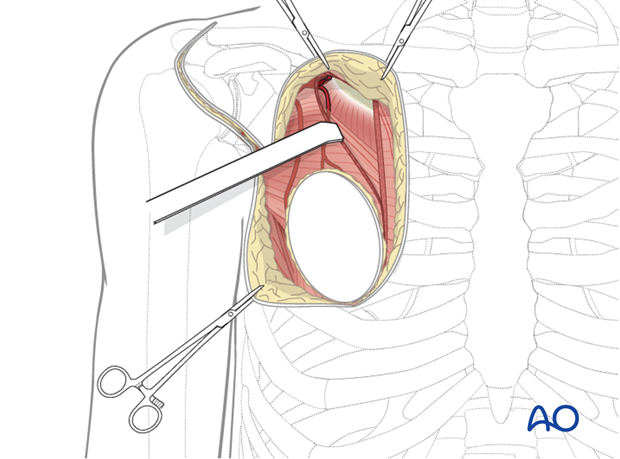
A subcutaneous tunnel is developed between the donor and the recipient sites, superficial to the clavicle. The pedicle can then be passed.
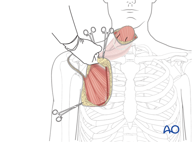
Now, the flap is ready to be passed through the tunnel and transposed into defect region.
Note: Careful handling of the skin paddle during this step avoids shearing of the skin perforators. Furthermore the pedicle must be inspected after the transposistion of the flap to ensure that it is not kinked.
Closure
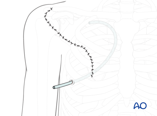
The chest wall is undermined and the incision is closed in layers.
A suction drain is used to drain the surgical site and prevent the formation of a hematoma or late seroma.
