Anterolateral thigh flap harvest
1. Introduction
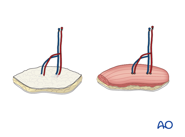
Both a free fascia, fasciacutaneous free flap and a myocutaneous free flap can be prepared from the anterolateral thigh.
Anatomy
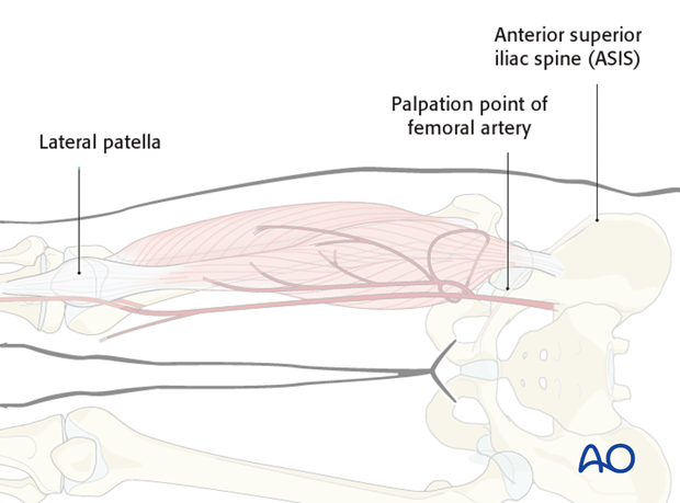
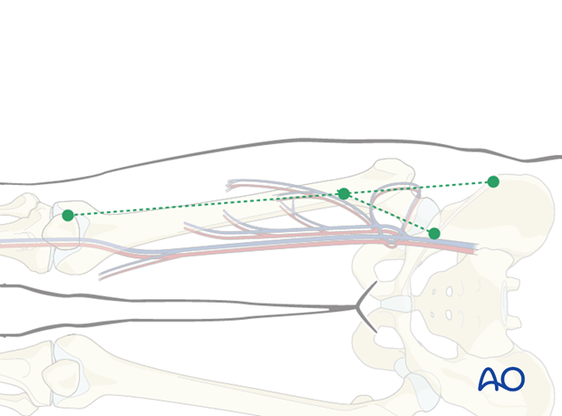
The following landmarks are identified:
- Anterior superior iliac spine (ASIS)
- Lateral patella
- Femoral artery (palpation for pulse)
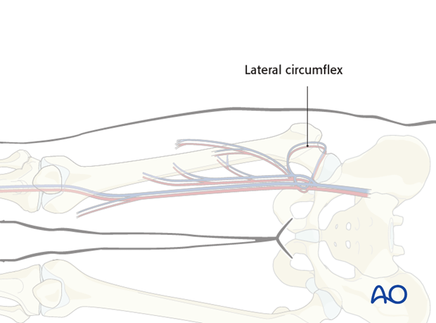
The vascular supply is based on the
Dominant: descending branch of lateral circumflex of the femoral artery and concomitant veins.
Alternative: oblique artery and concomitant vein
When a myocutaneous flap is needed, the vastus lateralis muscle is harvested together with the skin flap
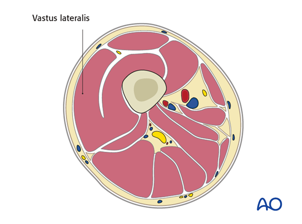
Flap design
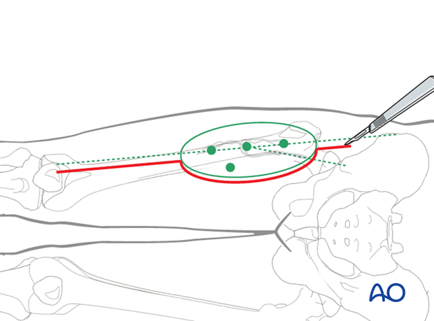
Two imaginary lines are drawn:
- from the ASIS to the lateral patella.
- from the center of the femoral artery running perpendicular to the inguinal ligaments to line 1
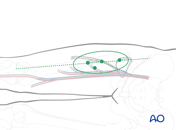
The crossing point between the two lines identifies the location of the (in most cases) most reliable and sizable perforator. The flap should be centered over this point as well as the first imaginary line.
The Dopplered mapping points should be incorporated in the flap design.
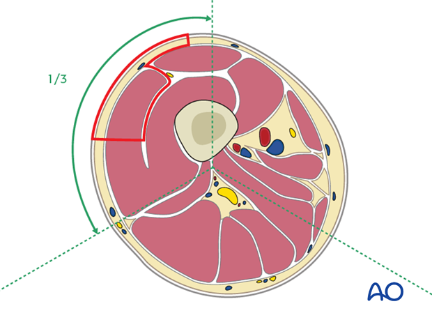
The width of the flap should not exceed 1/3 of the thigh circumferential. The length of the flap is only limited by the size of the vastus lateralis.
Primary closure for the maximum width can be felt with pinching test, if it exceed the limit, skin grafting might be required.
The flap design centred at septum between rectus femoralis and vastus lateralis muscles, width distribution shall be 1/3 medially (resting over rectus femoralis) and 2/3 laterally (resting over vastus lateralis muscle).
2. Anterolateral thigh free flap
Skin incision
The medial skin incision is made down through the fascia of rectus femoris and the lateral flap is raised in a subfascial plane until the muscular septum between rectus femoris and the vastus lateralis muscle is identified.
Dissection
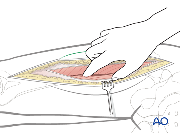
The incision is then carried through the fascia of rectus femoris and the lateral flap is raised in the subfacial plane using blunt dissection until the muscular septum between rectus femoris and the vastus lateralis muscle is identified.
Mobilization of pedicle
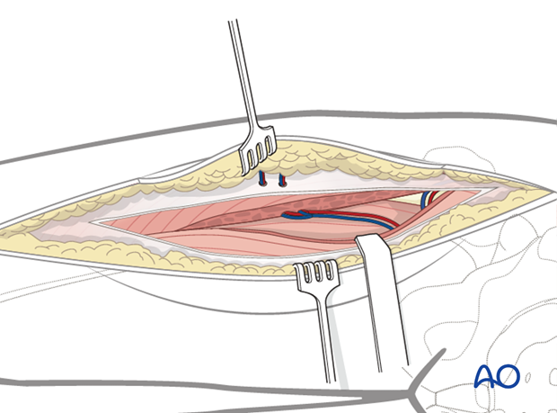
The rectus femoralis can be easily separated with digital dissection, and retracted medially and the pedicle is exposed above the vastus intermediate muscle.
The pedicle is mobilized until the bifurcation of the lateral circumflex femoral artery (LCFA).
Skeletonization of perforators
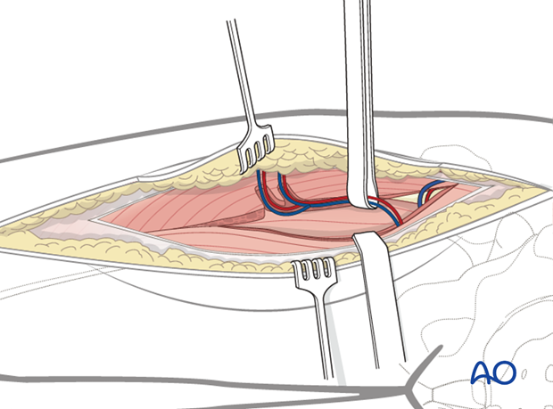
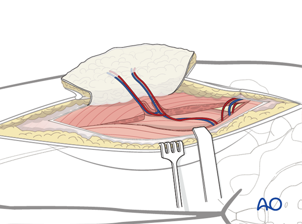
The perforators which start from the fascia lata and extend to the pedicle are skeletonized in retrograde motion. The perforators may be traveling through the septum (septocutaneous), however, more commonly vastus lateralis has to be dissected to isolate the musculocutaneous perforators.
Flap mobilization
The lateral incision is made down, and through the fascia lata and the flap can be mobilized. When the recipient site is ready, the pedicle is transected.
Closure
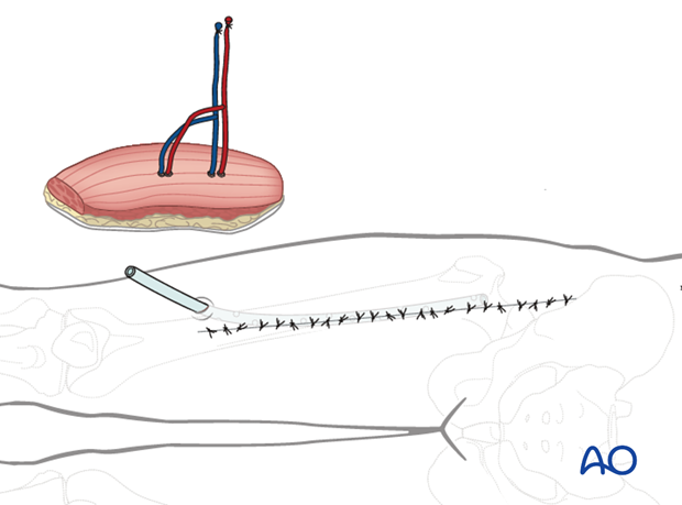
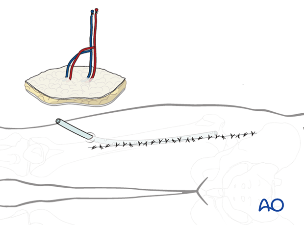
A drain is inserted and a primary wound closure performed.
3. Anterolateral thigh myocutaneous free flap
Skin incision
The medial skin incision is made down to the fascia lata and the skin flap raised laterally until the perforator is encountered.

Dissection
The muscular septum located between the rectus femoralis and the vastus lateralis muscles is identified.
The rectus femoralis is retracted medially and the pedicle is exposed by digital dissection. The pedicle is located above the vastus intermediate muscle.

Mobilization of pedicle
The pedicle is mobilized until the bifurcation of the lateral circumflex of the femoral artery.

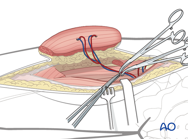
Flap mobilization
The lateral incision is made down through the fascia lata and the flap can be mobilized.
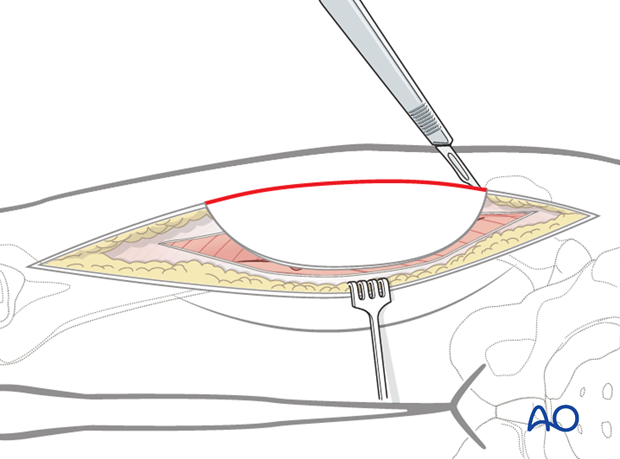
The flap is mobilized by transecting the vastus lateralis muscle. The amount of muscle that is harvested is dictated by the size of the defect.
Care has to be taken to include the perforators within the harvested muscle bundle.
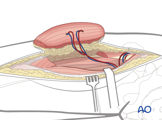
When the recipient site is ready, the pedicle is transected.
Closure
A drain is inserted and a primary wound closure performed.
