Transoral approach to the chin
1. Principles
Vestibular incisions
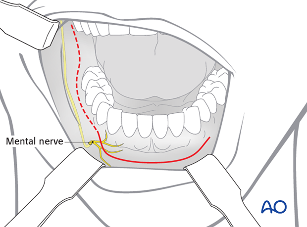
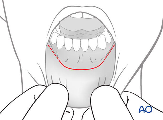
The transoral approach is the usual access for chin osteotomies.
The approach can be extended posteriorly (dashed line) in case a chin osteotomy is combined with an angle or ramus procedure.
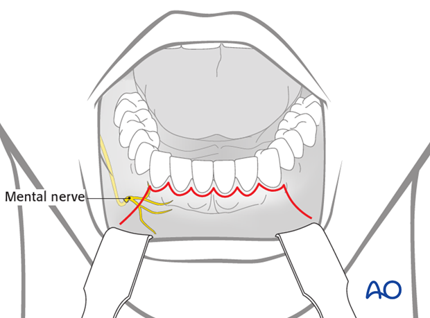
Alternatively a sulcular incision can be performed.
Neurovascular structures
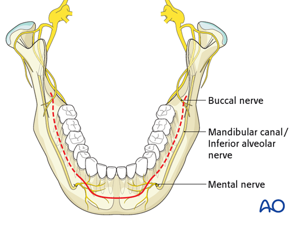
The mental nerve is a branch of the fifth cranial nerve (trigeminal nerve). This nerve provides sensation to the anterior mandibular vestibule, lip and chin.
When the incision is extended posterior to the canine teeth, the mental nerve can be damaged. Keep the incision superior to the mental nerve in the body region.
Particularly in the extended intraoral approach, care must be taken to protect the mental nerve in the anterior body region.
2. Intraoral incision
Mucosal incision
Unless contraindicated, infiltrate the area with a local anesthetic containing a vasoconstrictor.
Make an incision through the mucosa in the vestibule. Between the canines the incision is made 10–15 mm away from the attached gingiva in a curvilinear fashion. Posterior to the canine the incision is only 5 mm away from the attached gingiva, staying superior to the mental nerve.
Surgical flap dissection
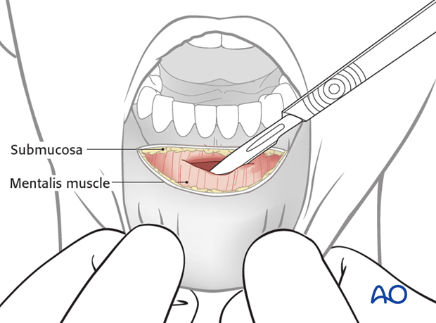
Incise the mobile mucosal layer lateral to the vestibular fold. Dissect a mucosal flap to expose the surface of the mentalis muscle. The branches of the mental nerve are located laterally just underneath the mucosal flap and must be avoided.
Mentalis muscle dissection
The mentalis muscle is divided near the alveolar bone ridge thus creating a stepwise incision. Later, during wound closure the mentalis muscle needs to be properly reattached to avoid a drooping chin.
Osteotomy site exposure
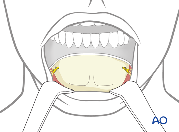
A mucoperiosteal flap is elevated to expose the chin down to the level of the lower border of the mandible. Great care must be taken to avoid stripping of the lingual periosteum. Lingual soft tissue attachment to the bone is needed to preserve blood supply. Both mental nerves are identified laterally; the subperiosteal dissection is continued underneath the nerves if needed.
Pearl: releasing the mental nerve (skeletonization)
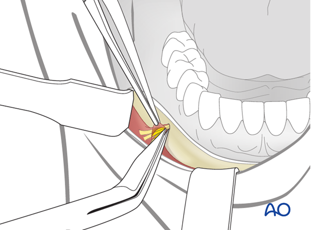
Skeletonization of the mental nerve allows for better soft-tissue retraction.
Fine scissors are used to cut the neurovascular bundle sleeve and spread parallel to the nerve. Scalpels could be used for that purpose but at higher risk of injuring the nerve.
3. Wound closure
After thoroughly irrigating the wound and checking for hemostasis the incision is closed. Anteriorly, the mentalis muscle is reapproximated with sutures to prevent drooping of the chin. The mucosa is closed with interrupted or running sutures.
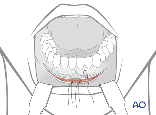
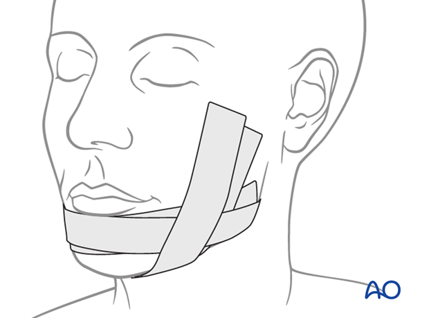
An elastic pressure dressing on the chin region helps support the soft tissues and prevent hematoma formation.
