Transcutaneous lower eyelid approaches
1. Principles
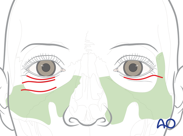
There are three basic transcutaneous approaches through the external skin of the lower eyelid to give access to the inferior, lower medial, and lateral aspects of the orbital cavity:
- Subciliary (A, synonym: lower blepharoplasty)
- Subtarsal (B, synonym: lower or mid-eyelid)
- Infraorbital (C, synonym: inferior orbital rim)
The subciliary approach can be extended laterally to gain access to the lateral orbital rim (D).
The course of the incisions is aligned to the slope of the natural skin creases, which become more apparent with age.
The skin of the eyelid is the thinnest in the human body. It has little or no dermis and almost no subdermal fat.
Hypertrophic scarring and keloid formation are very uncommon following lower lid skin incisions. In general, the scars become less conspicuous with time. The infraorbital incision can leave a permanent scar that can be prominent. The subciliary approach has the highest incidence of lid problems, such as ectropion.
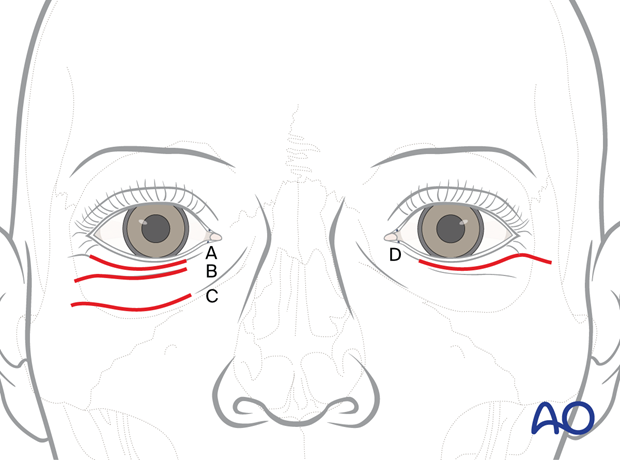
The approaches differ in the vertical position of the cutaneous incision lines. The path and length of dissection through the layers of the lower lid vary.
Due to the scarring and potential complications, the infraorbital approach has lost its former popularity. The infraorbital approach will not be detailed here.
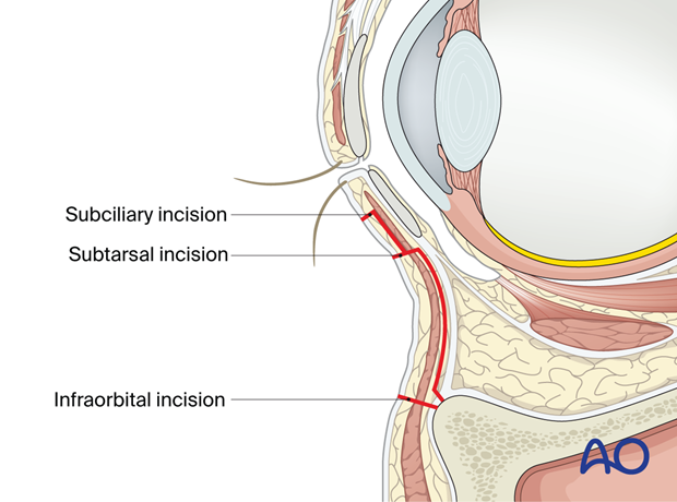
Access areas
Transcutaneous lower-eyelid approaches allow access to the lower region of the orbital cavity and upper midface.
The exposure of the infraorbital rim serves as the starting platform for the dissection of the periorbita.
The illustration outlines the approximate extent of the orbital bony surfaces exposed via the subciliary, subtarsal, and infraorbital approaches. There is little difference in the accessed areas along the floor, the medial and lateral rim, and the lower portions of the medial and lateral walls.
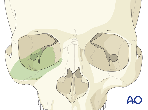
With a lateral extension of the subciliary incision and tissue traction, the lateral orbital wall and the zygomaticosphenoid suture become accessible.
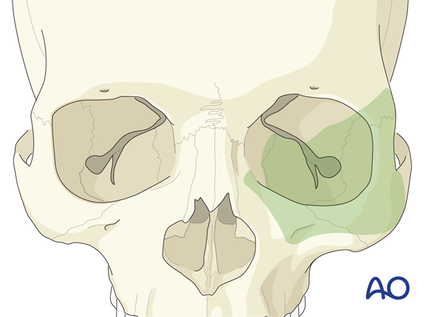
Exposure of midfacial skeleton
This illustration shows the area of bone exposure inferomedial of the infraorbital rim and across the zygomatic eminence, which can be reached following the release of the retaining ligaments and muscle attachments with subperiosteal dissection.
A connection with the surgical field through a maxillary vestibular approach will allow complete degloving of the lower and middle third of the midfacial skeleton.
2. Preparation: corneal protection
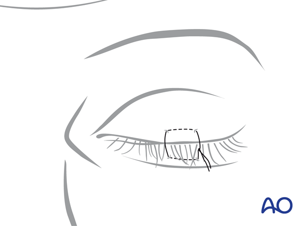
A temporary tarsorrhaphy is recommended for all three types of transcutaneous lower-eyelid incisions to help protect the cornea. This is done by employing a mattress suture.
In addition, you can use a corneal shield with eye ointment.
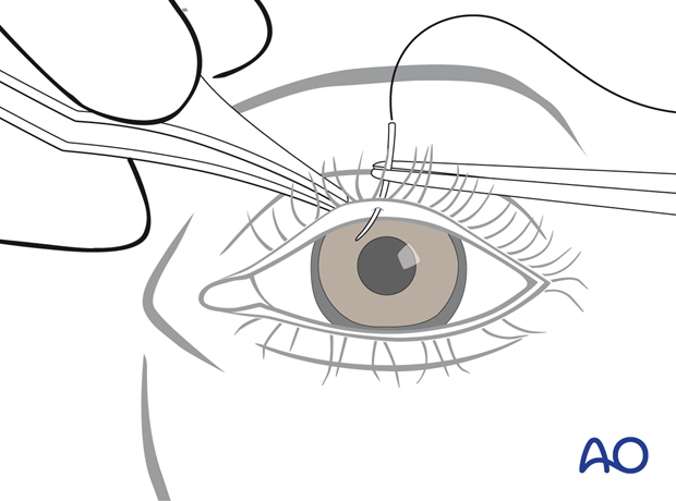
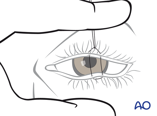
A 6.0 suture is passed through the skin of the upper eyelid and exits through the Gray line of the upper lid margin.
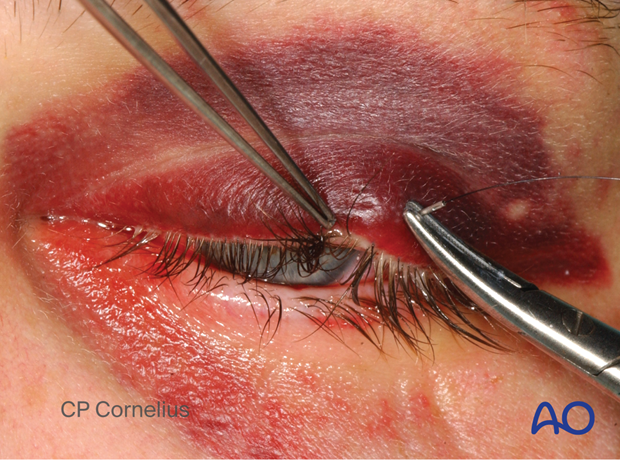
The needle is passed in the lower eyelid from the Gray line into the skin where it exits.
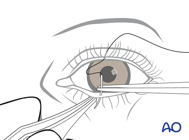
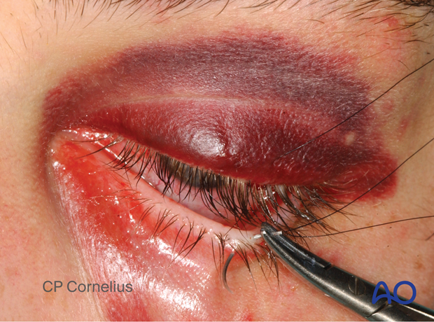
The suture is guided back, picking up the same soft-tissue portions in the lower and upper eyelids to complete the mattress loop.
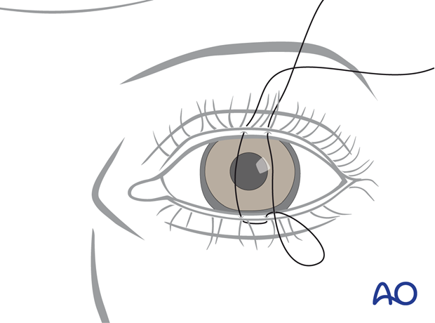
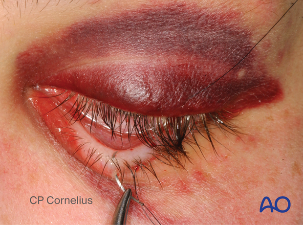
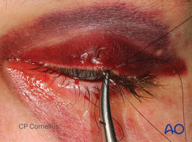
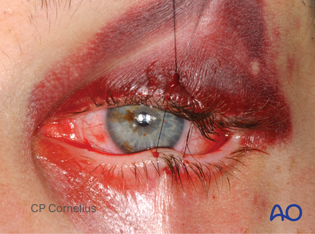
The tarsorrhaphy is not secured tightly, and some space is left between the knot and the upper-eyelid skin. A hemostatic clamp is used to grasp the suture and apply traction to the lower lid for full eyelid closure during the surgical procedure.
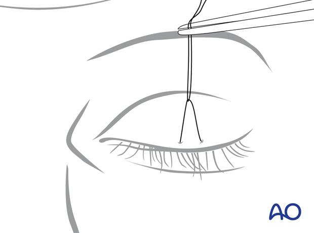
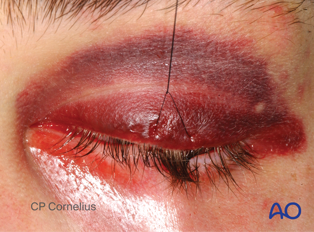
Since the suture was not fully tightened, when the hemostatic clamp is released, the lid may be opened for a forced duction test or evaluation of the pupil during the procedure.
3. Links to transcutaneous lower-eyelid approaches described
Click the following links to read a detailed description of the transcutaneous lower-eyelid approaches:
- Subciliary (A, synonym: lower blepharoplasty) including a lateral extension (D)
- Subtarsal (B, synonym: lower or mid-eyelid)


